


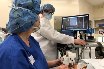
Researchers have developed and demonstrated the first handheld ophthalmology instrument with resolution-boosting adaptive optics technology that can image individual photoreceptors in the eye. The new portable instrument will allow improved diagnosis of eye diseases and could enable early detection of brain-related diseases and trauma.
In the Optical Society's journal Optica, the researchers report their new light-weight instrument measures just 10 x 5 x 14 centimeters. They say they tested the device in children and adults, demonstrating its ability to capture images of even the very small photoreceptors close to the center of the retina that play a key role in vision.
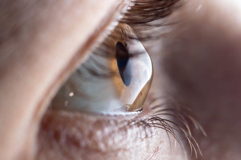
According to Optica, photoreceptors, specialised neurons that convert light entering the eye into signals sent to the brain, are the only neurons in the body that can be imaged non-invasively. It says imaging photoreceptors is not only important for diagnosing eye diseases but could also provide insights into processes occurring in the brain, adding preliminary studies have shown that changes in the retina can be observed during the early stages of diseases such as Alzheimer’s and after traumatic brain injuries such as concussions.
“Until now, the imaging systems required for high resolution photoreceptor imaging consisted of large, heavy components on an optical table that could only be used with cooperative adults sitting upright,” said research team leader Sina Farsiu from the biomedical engineering and ophthalmology departments at Duke University in the US. “Our portable handheld system could expand this important imaging technique to children and infants, as well as to adults who may not be able to sit upright and stare straight ahead.”
The system could be used on people who are in a reclined position as they undergo surgery, for example, Farsiu’s team reports. It could also help doctors rapidly assess possible brain trauma, such as in football players coming off the field with head injuries.
“Because of the limited resolution of MRI — the standard method for imaging the brain in living people— MRI-based assessment of disease or trauma to the brain cannot be done at the level of individual cells,” said Farsiu. “In the retina however, individual photoreceptors can be imaged at 100 times higher resolution than using brain imaging, allowing very subtle changes to be seen.”