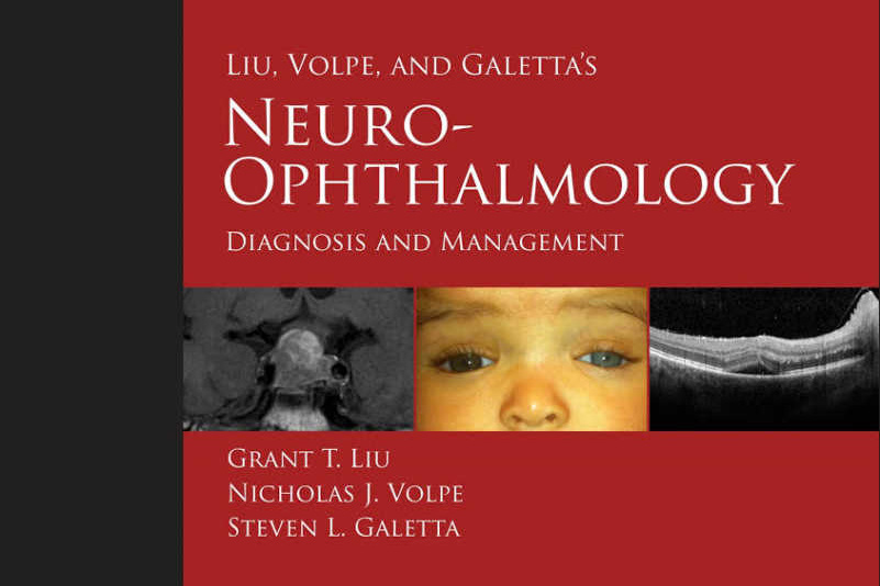
Book review: Liu, Volpe, and Galetta’s Neuro-Ophthalmology: Diagnosis and Management, 3rd Edition
Reviewed by Professor Helen Danesh-Meyer
This is a serious neuro-ophthalmology textbook. The authors cover diagnosis and treatment of neuro-ophthalmic conditions in one concise but dense textbook.
One of the strengths of the textbook is that it provides a large collection of photographs to illustrate the various conditions. Important information is highlighted in tables for easy access. It is written at the level of a really keen registrar and most of the information that one could need is in one textbook. Another important aspect of this book is that an e-version is available. By providing a personal code on the inside cover, the reader can access all the images and text. It is also associated with an extensive video collection which is particularly useful for examination of the efferent visual pathway and associated disorders: great for taking a saunter through on a rainy Friday night. In particular there is a huge collection of abnormalities for nystagmus that one does not see in clinics that often and a video is most certainly better than 1000 words in these circumstances. In addition, the authors often provide more than one image of a particular condition. This is very helpful for the reader to understand the range of potential presentations.
One of the weakness is the section which is focused on identifying the difference between glaucoma and non-glaucomatous optic neuropathy. There is an erroneous focus on ‘low tension glaucoma’ when in fact “ low” or “normal” tension glaucoma is merely a statistical construct with no strong pathological basis and there is no evidence that it is more likely for a patient to have a non-glaucomatous optic neuropathy with IOP <21 mmHg than in patients whose IOP is >21 mmHg.
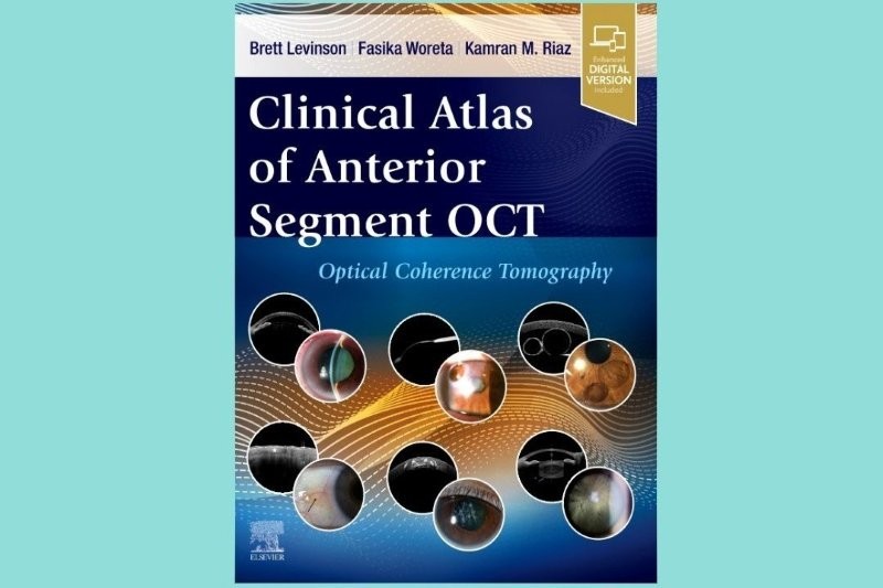
There are also some areas that would benefit from further detail. For example, there is a rapidly growing body of evidence discussing the difference between demyelinating optic neuritis, neuromyelitis optica, anti-myelin oligodendrocyte glycoprotein (anti-MOG) and chronic relapsing optic neuropathy. This is currently one of the most topical areas in neuro-ophthalmology and would have benefited from more discussion and detail. While the text does include numerous examples of new technologies such as optical coherence tomography (OCT), there are some notable exclusions. OCT has become quite an important part of neuro-ophthalmology and the usefulness of this tool is underestimated throughout the textbook. There are some examples of OCT in the differentiation of very obvious retinal disorders such as macular holes, but examples such as en face appearance of the macula in acute macular neuroretinopathy are not included and the importance of OCT in predicting visual outcome following surgery for chiasmal compression is omitted all together.
One of the sections that is very useful are the video clips on the various examination techniques. This allows the reader to observe neuro-ophthalmologists performing various aspects of the clinical examination and may provide an alternative perspective.
Overall, I can give this book a strong recommendation: it is easy to navigate, well-written by exceptional neuro-ophthalmologists, and comprehensive. It is an excellent foundation textbook providing middle ground between an encyclopaedic source and an atlas.
Professor Helen Danesh-Meyer is the head of Academic Glaucoma and Neuro-ophthalmology in the Department of Ophthalmology, University of Auckland.


