


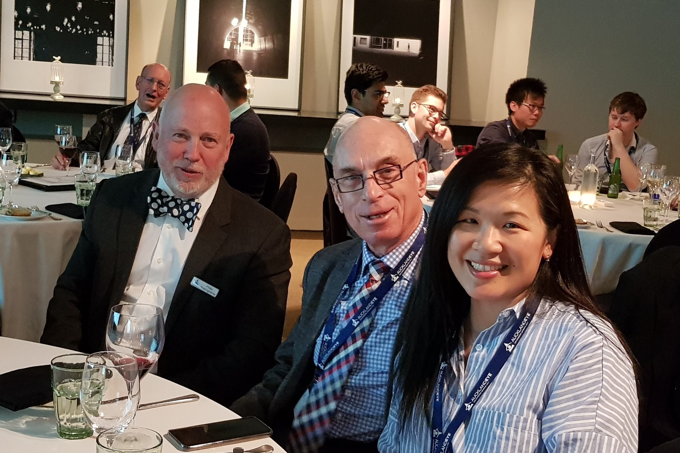
Auckland Eye celebrated 12 years of offering continuing education with the last of its Insight seminars for the year at the beautiful Orakei Bay venue in Auckland.
As the formal part of the evening got underway, we were treated to a moment of levity as optometrist Nigel Somerville was called to the podium to present his World Masters football team mate and Auckland Eye partner, Dr Paul Rosser, with his well-earned trophy shirt, celebrating their team’s silver medal win in the over 50s division.
Back to the education and Auckland Eye’s newest recruit, Dr Shenton Chew, opened proceedings with a presentation on the art and science of gonioscopy to aid understanding of when a narrow drainage angle needs referral and when the best management may involve surgery, particularly non-routine cataract surgery.
His opening statement that primary angle closure glaucoma (PACG) is three times more likely to lead to blindness than primary open angle glaucoma (POAG) and only gonioscopy can tell you if what you have in front of you is POAG or a PACG suspect, was an eye opener for many of us. Though every gonioscopist will have their personal favourite instrument, Dr Chew’s recommendation is the Goldman-style high-magnification lens which gives a high quality detailed image. He recommended having the room illumination as low as possible, as those most at risk of PACG can best be identified when the pupil is dilated in darkened conditions. Build up experience using it on everyone until the once difficult “squeezy” patient becomes easier and orientation in what was previously foreign territory becomes routine, he said.
The main course was served up by Associate Professor Phillip Polkinghorne, who said that 50% of us will experience a posterior vitreous detachment (PVD) once we are over 60. PVD is preceded by the development of large, optically-vacant spaces in the vitreous and an increase in floaters. Myopes and those who have had cataract surgery at an early age will experience PVD, and the consequent potential for retinal detachment and macular holes, earlier, with approximately 50% having a PVD within a year of surgery. He said a study is currently underway to determine whether oxidative damage caused by cataract surgery leads to vitreous detachment.
A/Prof Polkinghorne took us through the unique features of optic nerve head haemorrhages resulting from PVD. These are relatively common, occurring in 6% of PVDs and often confused with haemorrhages due to glaucoma. The location of the haemorrhage within the optic nerve head is related to the traction and can be preretinal, in the line of the retina or subretinal. Large population studies have shown more than 80% of people with optic disc haemorrhages did not have glaucoma. Further studies are pending.
The final course focused on staphylococcal marginal keratitis versus peripheral ulcerative keratitis, delicately served by Dr David Pendergrast. Differential diagnosis is complex as they may have similar signs and symptoms particularly on first presentation, but may come down to the responsiveness or not to topical therapy. Prevention via blepharitis treatment is always preferable, he said.
I, for one, am looking forward to more delectable offerings next year.