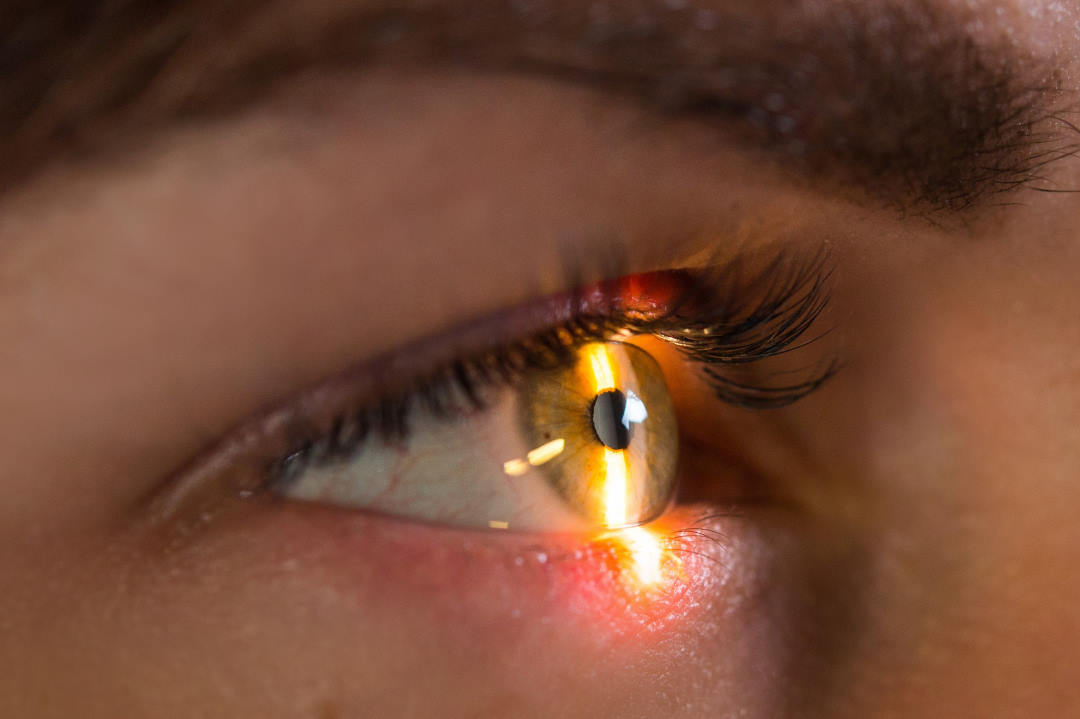
Glaucoma up close
Using methods originally developed by astronomers to view stars more clearly through Earth's atmosphere, Indiana University researchers have taken the first undistorted images of the eye’s trabecular meshwork affected by glaucoma.
Viewing this structure clearly for the first time should help improve glaucoma treatments, said Dr Brett King, head of advanced ocular care services and associate clinical professor at the IU School of Optometry. “Alterations of the trabecular meshwork, which allows fluid to drain, elevates pressure in the eye, leading to glaucoma. The problem is the meshwork can only be seen poorly with normal instruments due to its location, where the iris inserts into the wall of the eye, as well as the near-total reflection that occurs when looking through the cornea.
“The result of this low visibility is a lack of understanding about why age appears to cause the trabecular meshwork to function poorly. It also makes it difficult to study why certain glaucoma treatments that target the trabecular meshwork, such as laser therapies or invasive surgical procedures, fail while others succeed.”
To view the trabecular meshwork, IU researchers modified an existing ophthalmic laser microscope with a programmable mirror to correct for the eye's imperfections. Published in the journal of Translational Vision Science and Technology, study authors reported the technology, called "adaptive optics," was successfully used to image the trabecular meshwork in nine study participants, including two with pigment dispersion syndrome, an eye disorder that can lead to a form of glaucoma.
"Thanks to this research, the ocular drainage area of the eye can now be seen with much-improved clarity, which will improve our understanding of how this essential drainage area is being altered or damaged with age," said Dr King.


