


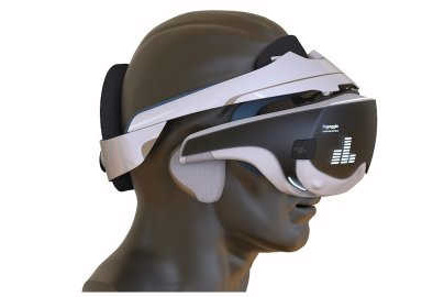
A new portable brain-computer interface, nGoggle, is proving promising for assessing visual function loss in glaucoma patients, said co-inventor Professor Felipe Medeiros from Duke University, North Carolina in an article in Ophthalmology Times.
The interface, developed by Prof Medeiros and colleagues, assesses visual function loss using multifocal steady-state visual evoked potentials (mfSSVEPs) associated with visual field stimulation.
The head-mounted unit integrates electroencephalography (EEG) with a cell phone-based display that appears in virtual reality-type goggles. The EEG uses dry electrodes positioned over the occipital region, and the goggles incorporate foam-based electro-oculogram sensors to assess eye movements, recognizing that fixation loss could affect the test results.
“Our brain-based objective method is completely portable and transmits data wirelessly through the network to an operating tablet or directly to the cloud via Bluetooth or Wi-Fi,” said Prof Medeiros.
In initial testing, nGoggle demonstrated greater accuracy compared with standard automated perimetry for discriminating eyes with glaucomatous optic neuropathy versus healthy eyes and was able to detect damage in eyes with preperimetric glaucoma. The evaluation was performed using a prototype device in a case-controlled study including 62 eyes of 33 glaucoma patients and 30 eyes of 17 healthy individuals. Glaucoma patients had relatively mild disease (average mean deviation ~–4 dB), and 11 of the 62 glaucomatous eyes had preperimetric disease. For the testing, the field of view was divided into 20 sectors, each flickering at a specific frequency. Global and sectoral mfSSVEPs were compared with global and sectoral SAP parameters.
Results showed the area under the receiver operating characteristic (ROC) curve for the brain-computer interface was 0.924, and it was significantly larger (better performance) than the SAP parameters used as comparators (mean deviation, 0.81; mean sensitivity, 0.80; pattern standard deviation, 0.77), Prof Medeiros noted.
Although eyes with preperimetric glaucoma had no evidence of visual function loss on SAP, they exhibited decreased mfSSVEP responses compared with healthy controls (0.280 versus 0.334), said Prof Medeiros. Sectors of visual function loss with the device corresponded with those identified on SAP, while thinner measurements on spectral domain-OCT also corresponded to lower amplitudes on the mfSSVEP, he added.
The technology has potential applications beyond assessment of glaucomatous visual function loss, said Prof Medeiros. “The device could potentially be used for assessing higher cognitive functioning by creating virtual reality tasks and monitoring brain activity through EEG.” The team is currently investigating its potential for longitudinal monitoring of glaucoma damage.