Insight into early AMD
The Centre for Eye Health Research in Sydney, Australia, says it has identified a potential marker for predicting drusen emergence for AMD prognostic and intervention studies.
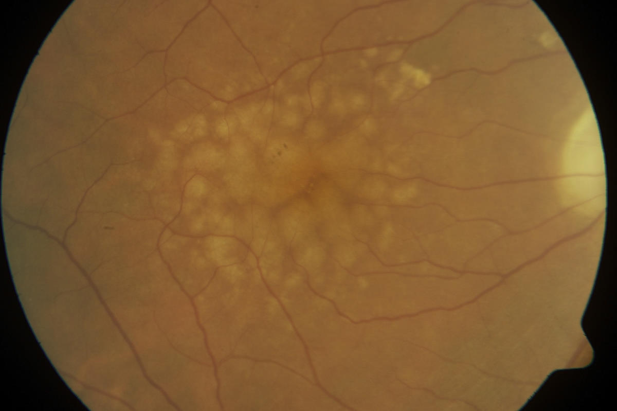
Drusen are associated with retinal thinning in age-related macular degeneration (AMD). These changes, however, have mostly been examined at single time points, ignoring the evolution of drusen from emergence to regression. Understanding the full breadth of retinal changes associated with drusen will improve understanding of disease pathogenesis, the centre says.
The study looked at how the natural history of drusen affects retinal thickness, focusing on the photoreceptor and retinal pigment epithelium (RPE) layers.
Spectral domain optical coherence tomography of subjects with intermediate AMD (n = 50) who attended the Centre for Eye Health for two separate visits (476 ± 16 days between visits) was extracted. Scans were automatically segmented with manufacturer software then assessed for drusen that had emerged, grown, or regressed between visits. For each identified lesion, the thickness of each retinal layer at the drusen peak and at adjacent drusen-free areas (150 μm nasal and temporal to the druse) was compared between visits.
Before drusen emergence, the RPE was significantly thicker at the drusen site (14.2 ± 2.6%) compared with neighboring drusen-free areas. There was a 71% sensitivity of RPE thickening predicting drusen emergence. Once drusen emerged, significant thinning of all outer retinal layers was observed, consistent with previous studies. Drusen growth was significantly correlated with thinning of the outer retina (r = −0.38, P < .001). Drusen regression resulted in outer retinal layers returning to thicknesses not significantly different from baseline.
Researchers concluded the natural history of drusen is associated with RPE thickening before drusen emergence, thinning of the outer nuclear layer as well as photoreceptor and RPE layers proportional to drusen growth, and return to baseline thickness after drusen regression. These findings could provide a potential marker for predicting drusen emergence for AMD prognostic and intervention studies and highlight that areas of normal retinal thickness in AMD may be former sites of regressed drusen.


