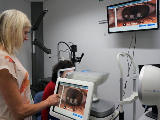
As the major cause of evaporative dry eye disease¹, meibomian gland dysfunction (MGD) requires careful assessment in the clinical setting. Morphological characteristics of meibomian glands are evaluated by meibography. This technique yields information on MG loss by observing the glands under infra-red illumination as light structures against a darker background during lid eversion (Fig 1)². The images are then subjectively graded by clinicians to indicate the severity of MG drop out, which is clinically important in the diagnosis and prognosis of MGD. However, test-retest and inter-observer variabilities in MG atrophy grading exist³.
Convolutional neural networks (CNNs) have shown promise in both classification and segmentation problems in ophthalmic imaging⁴. It is of interest to use deep learning methods to automate the process of evaluating atrophy in meibography images. Specifically, clinicians can use such methods to automatically segment the eyelids and identify areas of gland coverage in meibography images and then compute the amount of MG drop out.
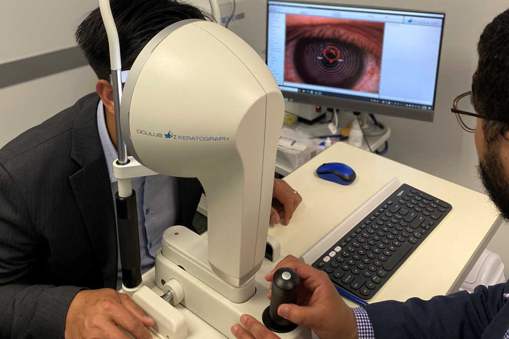
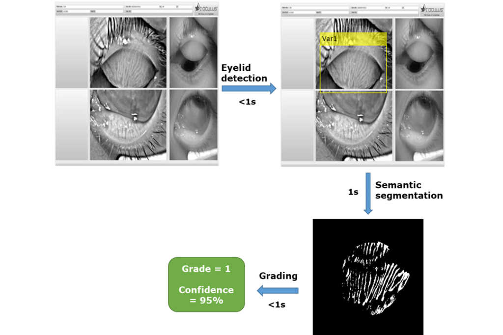
Our pilot study involves looking at a set of 821 upper eyelid images that were collected using the Keratograph 5M (Oculus) and clinically graded with the Pult score grading system. We have trained an artificial intelligence (AI) system on this data, such that we can now accurately distinguish the gland area and highlight the drop out region for the clinician and replicate the clinical score, all within 10 seconds of image acquisition.
Telehealth and AI are the fastest growing health-related fields in the post Covid-19 era. Ophthalmology and optometry have been at the forefront of smart technology adoption in the past decade⁵. AI enables automated and standardised clinical screening to be delivered across the nation with minimal clinical load. Combining forces between the Department of Ophthalmology and the School of Optometry and Vision Science at the University of Auckland, our group is aiming to develop an accurate AI algorithm for MGD screening, diagnosis and grading.
References
1. Lemp MA, Crews LA, Bron AJ, Foulks GN, Sullivan BD. Distribution of aqueous-deficient and evaporative dry eye in a clinic-based patient cohort: a retrospective study. Cornea. 2012; 31(5):472-478. doi:10.1097/ICO.0b013e318225415a
2. Fineide F, Arita R, Utheim TP. The role of meibography in ocular surface diagnostics: A review. Ocul Surf. Published online May 19, 2020. doi:10.1016/j.jtos.2020.05.004
3. Arita R, Minoura I, Morishige N, et al. Development of Definitive and Reliable Grading Scales for Meibomian Gland Dysfunction. Am J Ophthalmol. 2016; 169:125-137. doi:10.1016/j.ajo.2016.06.025
4. Vaghefi E, Hill S, Kersten HM, Squirrell D. Multimodal Retinal Image Analysis via Deep Learning for the Diagnosis of Intermediate Dry Age-Related Macular Degeneration: A Feasibility Study. J Ophthalmol. 2020; 2020:7493419. doi:10.1155/2020/7493419
5. Ting DSW, Pasquale LR, Peng L, et al. Artificial intelligence and deep learning in ophthalmology. Br J Ophthalmol. 2019; 103(2):167-175. doi:10.1136/bjophthalmol-2018-313173
Study lead and PhD student, optometrist Samira Hassanzadeh is a recent visiting scholar in the Department of Ophthalmology at Auckland University and holds a masters in medical education from Mashad University
Dr Eshan Vaghefi is a research fellow with the Molecular Vision Lab and Auckland Bioengineering Institute and a senior lecturer in physiological optics at the School of Optometry and Vision Sciences at the University of Auckland. He was responsible for training the AI system for this study.