Prof shrinks retina scanner
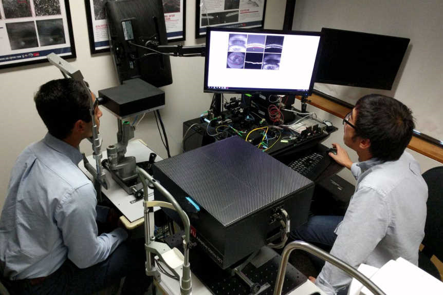
Engineering science professor Marinko Sarunic from Simon Fraser University, Canada has developed a new, shoebox-sized, retinal imaging scanner, that can still produce high-resolution, 3-D, cross-sectional retina, including individual photoreceptors, fine capillaries and blood vessels.
It’s unique small size makes it perfectly suited for everyday use in medical clinics and hospitals, said Prof Sarunic. “It’s a breakthrough in clinical diagnostics. With the high-resolution scanner, ophthalmologists and optometrists can detect damage and changes to small numbers of individual photoreceptors, giving them a diagnosis before the patient loses vision, and the potential to take preventative measures.”
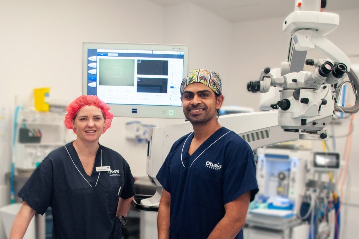
Dr Eduardo Navajas, a vitreoretinal specialist, says the scanner eliminates the need for dye injections, which are currently used to diagnose and monitor eye diseases like diabetic retinopathy and wet-AMD. “Early detection of abnormal blood vessels caused by wet-AMD and diabetes is essential to saving a patient’s vision. Dr Sarunic’s new imaging technology is benefiting patients, allowing us to diagnose and treat wet-AMD and diabetic eye disease before patients develop bleeding and permanent damage to their retina.”


