Residual refractive error after cataract surgery
Cataract surgery is the most commonly performed operation globally, with over 30,000 performed per annum in New Zealand. The surgery has transformed into a refractive procedure in recent years, with patients often expecting the surgery to minimise their dependence on glasses. However, residual refractive error following surgery is still relatively common. In 2015, postoperative visual and refractive outcomes in New Zealand were found to be comparable to international standards¹, which aims to have 85% of patients achieve a spherical equivalent (SE) within 1 dioptre (D) and 55% of patients within 0.5D of the intended target². This translates into more than 4,500 patients a year with a residual refractive error of >1D following surgery every year.
Multidisciplinary care of the cataract patient is continuously evolving to meet postoperative refractive benchmarks, with the development of an assortment of intraocular lenses (IOLs), advanced biometric measurements, modern IOL calculation formulae and the improvement in surgical options available to perform postoperative enhancement.
Current cataract surgery techniques
Phacoemulsification remains the gold standard technique for cataract surgery. It has replaced previously conventional methods such as intracapsular cataract extraction (ICCE), extracapsular cataract extraction (ECCE) and manual small incision cataract surgery (MSICS), due to advantages in visual outcomes and a lower complication rate.
Recently, the femtosecond laser has been employed for use in cataract surgery due to theoretically higher precision in surgical incisions, more predictable capsulotomy and reduced ultrasound energy. There is a sparsity of large, long-term studies comparing femtosecond laser-assisted cataract surgery to phacoemulsification, however, at this stage, there is no compelling evidence to suggest significant differences in refractive or visual outcomes. One European registry study suggests that femtosecond laser-assisted cataract surgery has a similar intraoperative complication profile compared with phacoemulsification but a higher rate of postoperative refractive surprise due to corneal oedema and posterior capsule opacification.

Fig 1. Proliferation of residual lens epithelial cells causing posterior capsule opacification with pearl-like clusters (left), and anterior capsular contraction with circumferential fibrosis (right)
Minimising preoperative sources of error
Recognising potential sources of postoperative refractive error is instrumental in achieving desirable postoperative outcomes. In most cases, a detailed ocular history, thorough examination and appropriate imaging is adequate for detecting significant risk factors that converge to lead to inappropriate IOL power selection. Such risk factors include ocular surface disease (OSD), contact lens wear, prior refractive surgery, keratoconus, astigmatism and extremes of axial length (AL).
Ocular surface disease
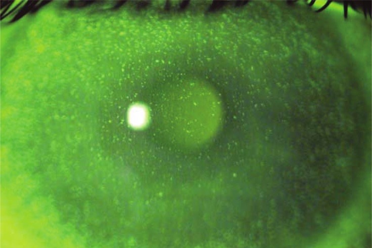
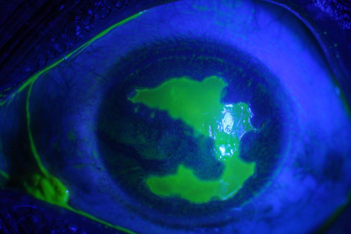
OSD is an underdiagnosed condition that has a prevalence of 5-50% globally. One study of patients presenting for cataract assessment revealed that 77% of cases exhibited corneal fluorescein staining, while only 22% had a formal diagnosis of dry eye. Untreated OSD resulting in hyperosmolarity and tear film instability, compromises the quality of the refractive surface, which directly affects keratometric measurements and can lead to erroneous measurements of keratometric astigmatism. Errors in measured astigmatism can be significant in IOL power calculations, particularly for astigmatic correcting (toric) IOLs.
Error due to OSD can be avoided through vigilant assessment and subsequent comprehensive treatment of the ocular surface for at least four weeks, at which point repeat biometry measurements will be necessary. Treatment should involve frequent lubrication and lid hygiene with adjuvant treatment such as topical steroid and oral azithromycin reserved for recalcitrant cases.
Contact lens wear
Contact lens wearers are also subject to an increased risk of postoperative refractive error. Extensive contact lens wear leading to prolonged hypoxia and subsequent corneal warpage can influence the accuracy of keratometric measurements³. Discontinuation of contact lens wear for a minimum of two to three days for soft lens and one to two weeks for rigid gas permeable lenses, prior to biometric and topographic measurements, is essential. A more prolonged discontinuation period may be required if the preoperative biometric and topographic measurements still exhibit evidence of corneal warpage, and multiple visits and measurements may be required in a small proportion of patients.
Refractive surgery
Prior refractive surgery, such as laser in situ keratomileusis (LASIK), laser epithelial keratomileusis (LASEK), photorefractive keratectomy (PRK) and radial keratotomy (RK), can instigate multiple errors in IOL power calculations due to erroneous preoperative keratometric measurements⁴. Traditional topography and biometry devices only measured the anterior radius of curvature while assumptions were made about the posterior cornea contribution for the index of refraction. After all types of refractive surgery, with the exception of RK where anterior-posterior changes are relatively proportional, these assumptions are violated. If not accounted for, patients with myopic refractive surgery generally experience a hyperopic shift following cataract surgery and a myopic surprise can be expected after hyperopic refractive surgery. Development of multiple modern IOL formulae and supplementary adjustment tools have tailored calculations to account for these factors, but it remains prudent upon the referring optometrist and the cataract surgeon to actively ask for a history of refractive surgery.
Keratoconus
Other conditions resulting in corneal irregularities, such as corneal ectasia, present similar challenges in IOL calculations. Accurate biometry measurements are notably difficult to obtain in patients with keratoconus, especially in those with severe disease who are more prone to cataract formation due to concurrent atopy and possible previous topical steroid use. Displacement of the steep axis causing misalignment with the visual axis and the presence of severe corneal distortion induces error in keratometric measurements and effective lens position, typically resulting in an underestimated IOL power and postoperative hyperopia. In preoperative assessments, vigilant detection for keratoconus, accurate staging and obtaining an understanding of the patient’s disease status and possible progression can be essential in minimising postoperative refractive error which may have accentuated downstream effects due to future interventions that may be required for the treatment of the ectatic process (eg. keratoplasty).
Astigmatism
Astigmatism can be challenging to address and accurate assessment of its magnitude is helpful in surgical planning. Previous research has revealed the residual refractive error is more common in individuals with higher levels of preoperative astigmatism. Varying degrees of astigmatism are managed differently. Patients with <1D astigmatism are generally managed with a monofocal IOL in conjunction with a steep axis surgical incision, whereas patients with >1D often benefit from a toric IOL⁵. A recent meta-analysis showed that toric IOLs are superior in astigmatism correction compared to corneal incisions, but these lenses need to be placed accurately on the steep axis in order to effectively neutralise astigmatism⁶.
Axial length and modern formulae
Patients with extremes of AL are also more prone to refractive surprise. The use of modern biometry devices that use optical coherence tomography (OCT) to generate accurate AL measurements as well as modern formulae has led to a significant reduction in refractive error. The recently introduced formulae analyse multiple biometric parameters such as AL, keratometry, lens thickness, anterior chamber depth and white-to-white distance, and employ regression models or artificial intelligence to better predict the postoperative position of the IOL (effective lens position) and improve refractive outcomes. In one recent study, the refractive accuracy was found to be most challenging to achieve in short eyes <22.0mm⁷.
Surgical causes
Intraoperative errors such as inaccurate IOL insertion, IOL placement outside the capsular bag or complications such as posterior capsular rupture can also lead to a significant postoperative refractive surprise.
Capsular causes
Late refractive changes following cataract surgery are often related to anterior/posterior capsular opacification and contraction. Mean shifts in spherical equivalent of 0.7D can occur from day one to two months postoperatively. Use of the Nd:YAG laser to perform a posterior capsulotomy or relive anterior capsular contraction can be very efficacious in restoring the optimal IOL position and treat late refractive change after cataract surgery.
One of the less common causes of reduced visual acuity can be attributed to capsular block syndrome, which is a rare complication with a reported incidence of 0.7%. Retained viscoelastic or fluid behind the IOL can cause anterior IOL shift, resulting in a myopic shift. Conservative management can result in resolution for some patients presenting with this condition in the early postoperative stage but Nd:YAG capsulotomy may be required to restore optimal IOL position.
Postop refractive error assessment
Patients are most commonly asked to see their optometrist four to six weeks following surgery to update their glasses as refractive errors generally stabilise between two to four weeks postoperatively⁸. When assessing refraction after cataract surgery, the use of subjective refraction is vital as automated refraction can be prone to error, particularly if a premium lens, such as a multifocal or extended depth of focus IOL, has been implanted. A thorough anterior and posterior segment examination can help to identify the causes of refractive error.
Postop refractive error correction
Patients should be reassured regarding enhancement options for residual refractive error correction. Corrective procedures can be cornea-based with laser vision correction or lens-based with secondary IOL implantation or IOL exchange.
Laser vision enhancement is the most common way of dealing with residual refractive error following cataract surgery. It is typically performed after a waiting period of at least six weeks but ideally three months following surgery. This allows for stabilisation of the refraction and also the surgeon to treat the ocular surface in patients who are often elderly and more at risk of OSD. During this period, the optometrist plays an extremely important role providing reassurance and temporary refractive correction to allow for functional vision. It is, however, necessary to exclude contraindications such as corneal pathology and extreme dry eye.
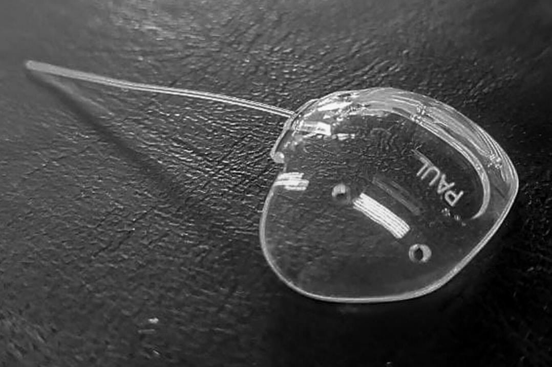
Lens-based correction may be preferred where the residual refractive error is large. IOL exchange can also be utilised to correct residual refractive errors but is often the last resort option as it is associated with a significant risk of intraoperative complications and, in most circumstances, secondary IOL insertion into the ciliary sulcus is a more favourable and safer treatment option.
One study found that LASIK enhancement was more efficacious and predictable compared to lens exchange and piggyback lens, with 92.85% of eyes achieving a SE within ±0.50D and 100% of eyes within ±1.00D⁹.
When counselling patients regarding postoperative residual refractive error, it is important to acknowledge the patient’s concern and ensure preoperative measurements are reviewed to confirm the correct IOL was implanted. After ensuring the correct IOL was implanted, it is also important to reiterate that residual refractive error is not an uncommon issue. Patient reassurance regarding the availability of solutions can eliminate potential distress and dissatisfaction, but before committing to an enhancement procedure, it is vital to ensure the patient understands the visual consequences, particularly if they are left myopic after surgery and are enjoying the benefits of unintended “monovision”.
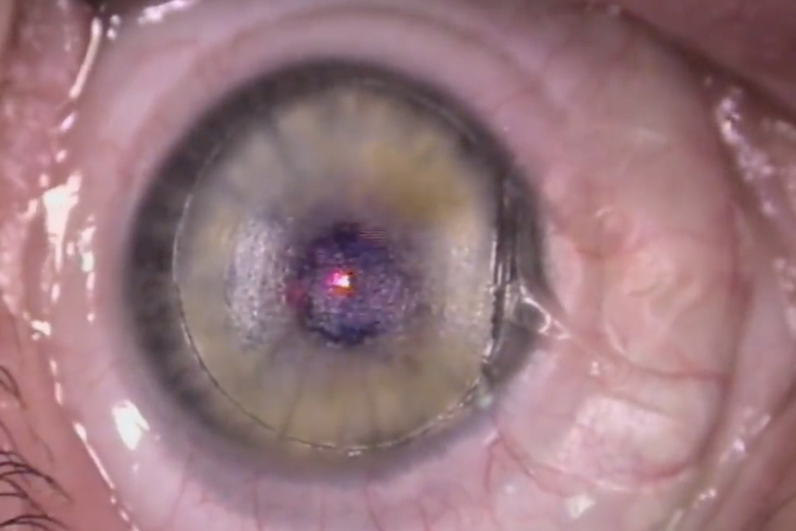
Fig 3. LASIK enhancement in a patient with hyperopic refractive surprise following routine cataract surgery
Conclusion
Advances in cataract surgery have enabled patients to achieve improved visual outcomes, with the rise of more IOL options, surgical accessories and improved precision in IOL power calculation10. Despite such developments, postoperative refractive error remains relatively common.
Refractive error after cataract surgery can be minimised through recognition and correction of potential risk factors. In instances where the amount of refractive error is perceived as unacceptable, enhancement procedures can provide excellent refractive and visual outcomes.
Whilst perfection in postoperative refraction cannot yet be guaranteed, achieving patient satisfaction is absolutely within reach through patient-centred care, appropriate counselling and adequate utilisation of enhancement procedures following cataract surgery.
References
- Kim BZ, Patel DV, McGhee CN. Auckland cataract study 2: clinical outcomes of phacoemulsification cataract surgery in a public teaching hospital. Clin Exp Ophthalmol. 2017;45(6):584-91
- Gale RP, Saldana M, Johnston RL, Zuberbuhler B, McKibbin M. Benchmark standards for refractive outcomes after NHS cataract surgery. Eye (Lond). 2009;23(1):149-52.
- Lewis JR, Knellinger AE, Mahmoud AM, Mauger TF. Effect of soft contact lenses on optical measurements of axial length and keratometry for biometry in eyes with corneal irregularities. Invest Ophthalmol Vis Sci. 2008;49(8):3371-8.
- Hoffer KJ. Intraocular lens power calculation after previous laser refractive surgery. J Cataract Refract Surg. 2009;35(4):759-65.
- Khan MI, Muhtaseb M. Prevalence of corneal astigmatism in patients having routine cataract surgery at a teaching hospital in the United Kingdom. J Cataract Refract Surg. 2011;37(10):1751-5.
- Lake JC, Victor G, Clare G, Porfírio GJ, Kernohan A, Evans JR. Toric intraocular lens versus limbal relaxing incisions for corneal astigmatism after phacoemulsification. Cochrane Database Syst Rev. 2019;12(12):Cd012801.
- Tang KS, Tran EM, Chen AJ, Rivera DR, Rivera JJ, Greenberg PB. Accuracy of biometric formulae for intraocular lens power calculation in a teaching hospital. Int J Ophthalmol. 2020;13(1):61-5.
- Caglar C, Batur M, Eser E, Demir H, Yaşar T. The Stabilization Time of Ocular Measurements after Cataract Surgery. Semin Ophthalmol. 2017;32(4):412-7..
- Fernández-Buenaga R, Alió JL, Pérez Ardoy AL, Quesada AL, Pinilla-Cortés L, Barraquer RI. Resolving refractive error after cataract surgery: IOL exchange, piggyback lens, or LASIK. J Refract Surg. 2013;29(10):676-83.
- de Silva SR, Evans JR, Kirthi V, Ziaei M, Leyland M. Multifocal versus monofocal intraocular lenses after cataract extraction. Cochrane Database Syst Rev. 2016;12:Cd003169.
Dr Ye Li is a PGY1 junior doctor with a strong interest in ophthalmology.
Dr Mo Ziaei is a senior lecturer at the University of Auckland, a cataract, cornea and anterior segment specialist, practising at Greenlane Clinical Centre and Re:Vision in Auckland.


