For the first time the World Glaucoma Congress (WGC) came to the Southern Hemisphere. With many of the big names in glaucoma making the trip downunder, this conference promised to be a great way of catching up with the latest thinking in glaucoma without having to cross too many time zones! It was also a good chance to see colleagues from around the globe and find out what has been happening in the latest round of professorial musical chairs (or maybe that should be game of thrones!).
New frontiers: diagnosis
Once I had managed to avoid getting sucked into the AusPack packaging conference being held in the other half of Melbourne’s massive convention centre, I made it to the WGC’s first-day main event: New Frontiers in Glaucoma, chaired by Professor Sir Peng Khaw.
This kicked off with a talk from Dr Naama Hammel, a glaucoma specialist who now works for Google on its artificial intelligence (AI) platforms, who helped us understand the difference between AI, machine learning and deep learning. There is now a programme that can teach a machine how to machine learn. Luckily (for the moment), it still needs humans to be involved to set the truths on which the machines can learn. Though this is proving to be the biggest challenge - getting glaucomatologists to agree on a diagnosis!
Another speaker highlighted future developments in OCT that may help in diagnosis, such as widefield imaging and analysis of lamina cribrosa changes. Functional measures using OCT angiography and integration with central visual field testing and imaging of apoptosing retinal ganglion cells are also on the horizon.
Another interesting topic was the role of genomics in targeting populations who may need more intensive screening for glaucoma. This may also allow us to predict which patients will respond best to different treatments.
Professor Keith Martin discussed the concept of the “sick” retinal ganglion cell (RGC) and how they may be damaged by different mechanisms, and his ongoing work on RGC mitochondrial transport that might lead to ways of “rescuing” sick RGCs.
New frontiers: medical management
On Thursday morning I had conference app apoplexy thanks to there being three things at once I had flagged as being of interest! I attended the New Frontiers in Medical Management session. This included a talk by Professor Robert Weinreb about using a compound which works on the nitric oxide signalling pathway to lower intraocular pressure (IOP) as nitric oxide shrinks trabecular meshwork cells. There is now an US Food and Drug Administration (FDA) approved drug, latanoprostene bunod (latanoprost attached to the nitric oxide signaller) which is effective at lowering IOP, more than latanoprost alone. This may also have a pressure independent effect on protecting RGCs.
Another interesting talk I attended, covered how existing drugs may be administered more reliably in different ways such as via contact lenses, punctal plugs, refillable slow release reservoirs or long-acting injections. Several of these new drug delivery methods have the potential to change the way we practice in the not too distant future.
An eye opening conclusion to this session was a talk on how meditation may help in glaucoma. The purported link was in elevated cortisol levels causing higher IOP. A randomised, controlled trial has shown a slight decrease in IOP in glaucoma patients who performed 45 minutes a day of meditation for six weeks. This also resulted in improved brain oxygenation and reduced stress markers in serum.
New frontiers: ocular imaging
Being a bit of an ocular imaging geek, I was looking forward to the session entitled, Seeing is believing – an update on ocular imaging.
This included a fascinating study on aqueous flow through the aqueous collector channels using both fluorescein and ICG to show the variability between patients and even the dynamic change within patients. This was then further explored by inserting iStent trabecular bypass devices into cadaver eyes and watching the change in flow from the collector channels, which raised the question of whether we should be trying to rescue areas of low flow or enhance areas that already drain well. This is still under investigation but may allow us to tailor treatment more precisely.
OCT angiography of the optic nerve head has been a technology looking for a purpose for some time. The latest buzzwords are ‘choroidal microvascular drop out’, which occurs in glaucoma patients and in some cases can be present before visual field changes. A major issue currently, is the degree of test-retest variability which may reduce its effectiveness for detecting change over time and it is still inferior to retinal nerve fibre layer (RNFL) measurements in diagnostic accuracy.
There was also a presentation on the challenges in imaging myopic eyes with the suggestion that ganglion cell analysis maybe more helpful than RNFL given the frequent situation of altered disc morphology causing issues with obtaining and interpreting optic disc scans. However, this is awaiting more work to establish useful normative databases for these eyes, but again there’s hope OCT angiography might provide additional diagnostic help.
New frontiers: tools and techniques
Friday morning kicked off with The glaucoma surgery armamentarium - what's in our 2019 bag of tricks.
This ran through the various minimally invasive glaucoma surgery (MIGS) options, some of which looked familiar but had changed names thanks to some corporate mergers and acquisitions in this ever evolving and potentially highly lucrative market. The most promising of these, however, is the PreserFlo (formerly known as the Innfocus microshunt, which was also formerly known as the MIDI Arrow!). This is a fine tube implant made of a biocompatible material that is inserted after conjunctival dissection, as created for a trabeculectomy but where one end is injected via a needle into the anterior chamber, so it’s actually a LIGS (less invasive glaucoma surgery) rather than MIGS.
This was followed by a spirited defence of trabeculectomy with the speaker pointing out the long-term track record of trabeculectomy compared with MIGS devices and studies appearing to indicate less complications than glaucoma tube implants. He pithily re-labelled MIGS as ‘maximal income glaucoma surgery’!
Back to clever glaucoma drainage devices, there is a new aqueous shunt called an Eyewatch, where the flow (and hence IOP) can be controlled externally by a special magnet, called the EyeWatch pen, which controls drainage to the EyePlate. This is a very attractive concept but still rather bulky in its current form.
In the session enticingly titled, New Potions from the Hogwarts Cauldron for Treating Glaucoma, Professor Jonathan Crowston attempted to put the record straight after some sensationalist media reporting of research suggesting that niacin (vitamin B3) may be the holy grail of glaucoma treatment.
The rational for B3 treatment is its role in the oxidative phosphorylation pathway that is impaired in many neurodegenerative diseases and that there is increasing evidence of mitochondrial function being reduced in retinal ganglion cells in glaucoma. Studies on mice who are prone to glaucoma had shown niacin to be remarkably effective in preventing the development of glaucoma and Professor Crowstons’ team has an interventional study underway to further assess its effectiveness in glaucoma patients.
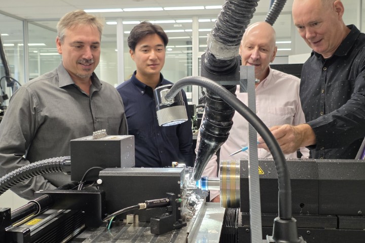
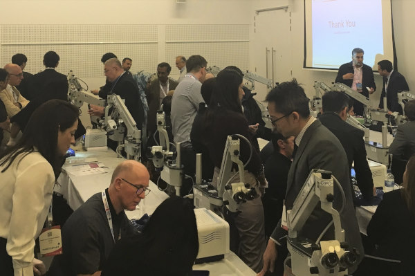
I also attended a wetlab on the use of the micropulse cylodiode laser. This is a new type of laser system that unlike traditional cyclodiode lasers (which aim to ablate the ciliary body) is aimed at both the pars plana and pars plicata to both reduce aqueous production and increase uveoscleral outflow.
Patient selection, pre- and post-op management were discussed. Then we had a chance to get a hands-on trial with the new laser probe and discuss tips on intra-operative techniques. It certainly appears to be a useful addition to our range of non-surgical options.
In conclusion
Overall this was a great conference with many stimulating sessions and chances to network with colleagues from around Australasia and the world. I will be looking forward to the next one in the similarly low jet lag location of Kyoto in 2021.
Dr Graham Reeves is a glaucoma specialist practicing at the Eye Institute and the Manukau SuperClinic in Auckland.