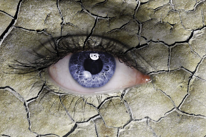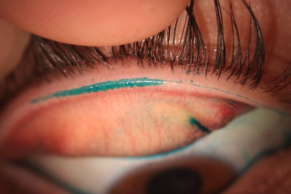
Clinically, this association appears to be reflected in “lid wiper epitheliopathy” (LWE), a staining pattern observed at the upper and/or lower lid margins following lissamine green instillation and lid eversion (Fig 1). But if vital stains are able to highlight a degree of cellular damage, what does this damage look like at a cellular level?
While dry eye and contact lens wear are recognised to be associated with cellular changes of the ocular surface (including within the cornea, limbus and bulbar conjunctiva), we know surprisingly little about clinically-relevant variations in the cellular anatomy and physiology of the lid wiper; the area in exclusive apposition with the eye’s surface during blinking. The cornea, bulbar or tarsal conjunctival are commonly and easily assessed by application and removal of a membrane to which superficial cells adhere. This “impression cytology” technique is a quick and convenient tool for sample collection from patient prior to histological cellular analysis. However, the narrow, sharply curved lid margin does not easily lend itself to such a sampling method. The optimisation of impression cytology for lid marginal use was the focus of my PhD studies at the University of Waterloo in Canada.
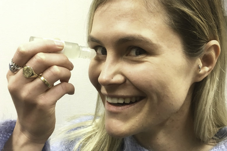
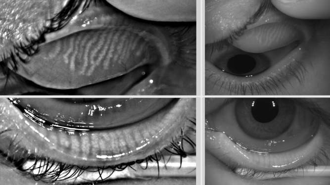
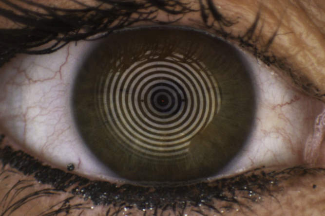
During this time, we determined the ideal membrane material, the optimal location, angle and pressure, and the duration of application (Fig 2). We chose specialised histological dyes that change their colour according to the keratinisation level of cells to reflect friction at the lid margin (Fig 3).
Fig 2. Impression cytology on the upper lid wiper region
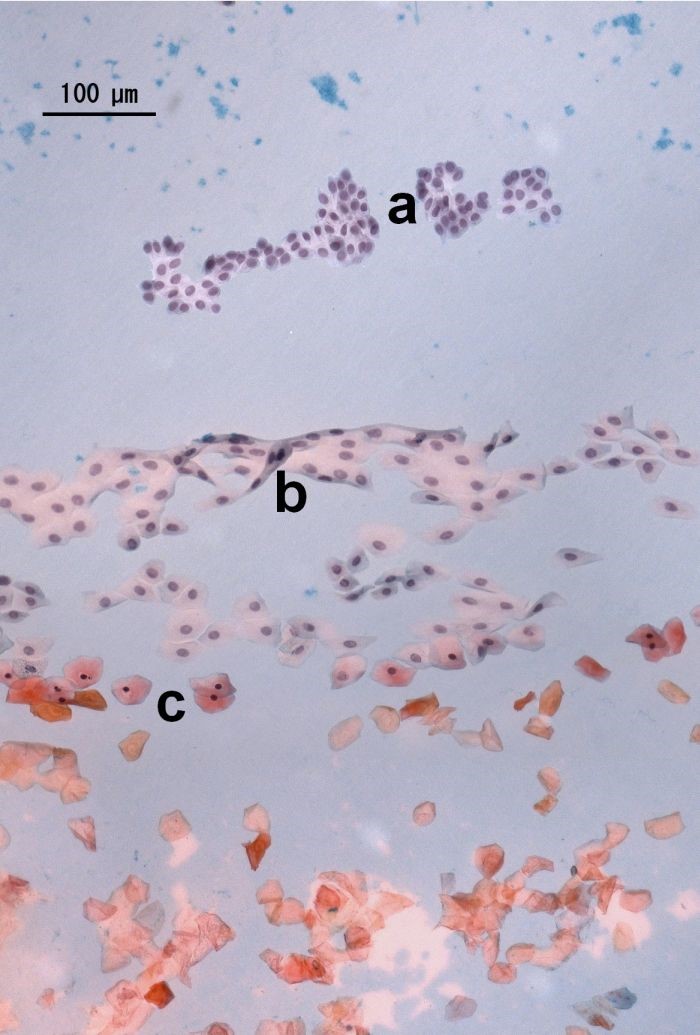
Fig 3. Cells of the lid wiper region show varying morphology and different keratinisation degrees through differential staining dyes: no keratinisation (a); advanced keratinisation (c); and incipient keratinisation (b)
Armed with this new tool, lid marginal epithelial cells were collected from patients presenting with varying levels of LWE, including contact lens wearers and non-lens wearers with a range of self-reported discomfort and dryness levels. By investigating the cellular morphology (size, shape, state, type, number of cells etc.) of the lid margin and its correlation with clinical signs and subjective symptoms, we are hoping to shed new light on the role of this region for dry eye and contact lens patients.
A video demonstrating this technique as well as our published methods paper are available at www.imuntz.com/ic. Stay tuned for a full report on these studies in an upcoming issue of NZ Optics!
Reference and pictures reproduced with permission, courtesy of A Muntz, K van Doorn, L N Subbaraman, L W Jones, Impression cytology of the lid wiper area, J Vis Exp. (2016) e54261–e54261.
Dr Alex Muntz, an optometrist, is a post-doctoral research fellow in the Ocular Surface Laboratory at the University of Auckland and was previously a clinical research scientist with the University of Waterloo’s Centre for Ocular Research & Education