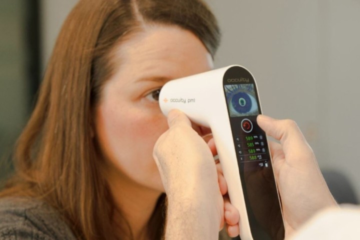
A review of the use of fluorescence lifetime imaging ophthalmoscopy (FLIO) has shown it offers exceptional detail that can aid early diagnoses and monitoring of retinal diseases.
Led by Dr Lydia Sauer from the University of Utah School of Medicine in the US, the review team compared FLIO with conventional autofluorescence intensity imaging. The latter relies on the fluorescence of lipofuscin, a fluorophore found in the retinal pigment epithelium. FLIO expands the level of information by capturing autofluorescence lifetimes.
Healthy eyes exhibit the same characteristic pattern of short FLIO lifetimes in the foveal centre and long lifetimes at the optic nerve, explained Dr Sauer. In contrast, eyes with retinal diseases are characterised by numerous different autofluorescence patterns, depending on the diseases present. FLIO is able to detect changes related to various retinal diseases, such as age-related macular degeneration (AMD), albinism, Alzheimer’s disease, diabetic retinopathy (DR), macular telangiectasia type 2, retinitis pigmentosa and Stargardt disease. Some of these changes can even be visualised in healthy eyes, possibly indicating a risk of developing such diseases, she said.
Sauer and her colleagues noted that FLIO also demonstrated that the geographic atrophy secondary to AMD has a markedly different appearance from the retinal atrophy associated with Stargardt disease, which is interesting since the two diseases can appear very similar when using autofluorescence intensity imaging.