Canada’s Photomedicine Labs at the University of Waterloo has demonstrated the first, non-contact, in-vivo imaging of ocular tissue using photoacoustic remote sensing (PARS) microscopy and swept-source optical coherence tomography (SS-OCT).
Although it could be another two years before clinical human trials begin, having demonstrated the technique’s application in mice, researchers said they anticipate it will allow measurement of chromophore concentration, melanin concentration and oxygen saturation, potentially providing far earlier and more accurate eye disease diagnosis. The loss of melanin in the retinal pigment epithelium is known to be a major factor in the progression of age-related macular degeneration (AMD), for example, while abnormal oxygen saturation is seen in diabetic retinopathy, glaucoma and retinal vein occlusion.
“For the first time, not just in ophthalmology, but in the entire medical field, diagnosis and treatment of disease could be made prior to structural change and functional loss,” explained Dr Richard Weinstein, co-founder of the university’s Ocular Health Centre in Nature.
Described as a non-contact version of photoacoustic microscopy (PAM), which can image any target that absorbs light energy, PARS paired with SS-OCT has the potential to provide chromophore selective absorption contrast in concert with depth-resolved scattering contrast in the ocular environment, said researchers. The study team is now planning a more comprehensive study.
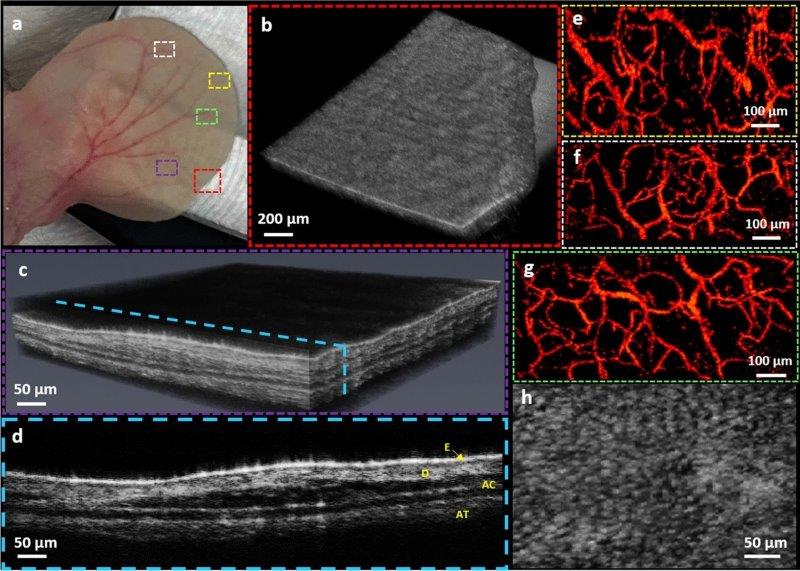
In-vivo imaging of mouse ear using multimodal PARS-OCT system. Corresponding white light photograph of the mouse ear, dotted rectangles indicate the PARS and OCT imaging regions (a). Volumetric rendering obtained by SS-OCT image (b). Volumetric OCT image showing different layers inside the ear tissue (c). Cross-sectional B-scan showing distinctive layers in the mouse ear tissue, E: Epidermis, D: Dermis, AC: Auricular cartilage, AT: Adipose Tissue (d). Vasculature of the ear obtained by PARS absorption mechanism (e–g). MAP image of the PARS scattering contrast showing epidermal ridges (h). Credit: PhotoMedicine Labs