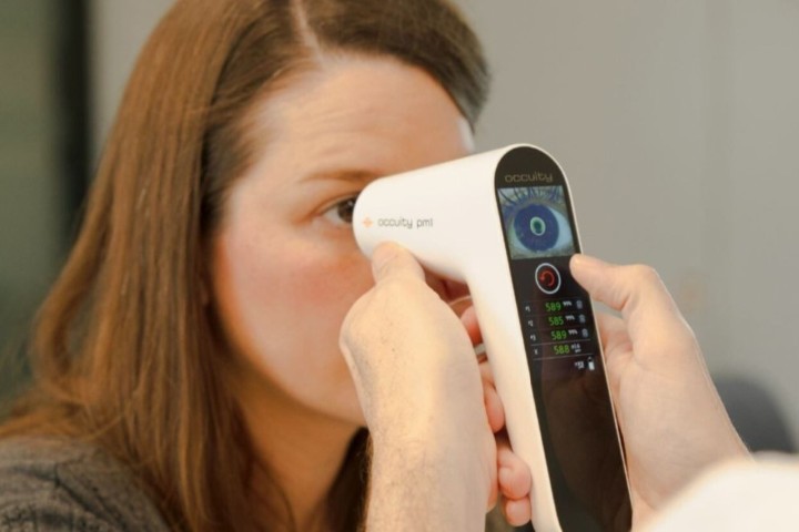


Researchers at the Swiss technical university École polytechnique fédérale de Lausanne (EPFL) have developed a prototype device which flags deterioration of retinal pigment epithelial (RPE) cells to diagnose degenerative eye disorders long before the onset of symptoms.
After capturing 100 raw images in less than five seconds, EPFL’s ‘Cellularis’ aligns and aggregates them to produce a single, high-quality picture, said researchers. Cellularis’ beams focus obliquely through the sclera, with light waves captured by the camera as they exit the through the pupil, said Associate Professor Christophe Moser, who heads EPFL’s Laboratory of Applied Photonics Devices. “This circumvents the problem (when using other imaging modalities) of excess light caused by the highly reflective cone photoreceptor cells when the retina is illuminated via the pupil.”
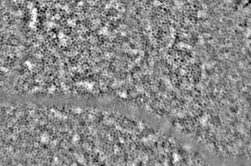
In early clinical trials with 29 healthy patients, researchers said the images generated were precise enough to quantify the morphological characteristics of the participants’ RPE cells. This, they said, is vital to making early diagnoses of conditions such as retinitis pigmentosa, diabetic retinopathy and age-related macular degeneration.