


US researchers using a mouse model of cerebrovascular disease found retinal vascular imaging and phenotype can function as a biomarker for Alzheimer's disease and related dementias (ADRD), particularly vascular contributions to cognitive impairment and dementia (VCID).
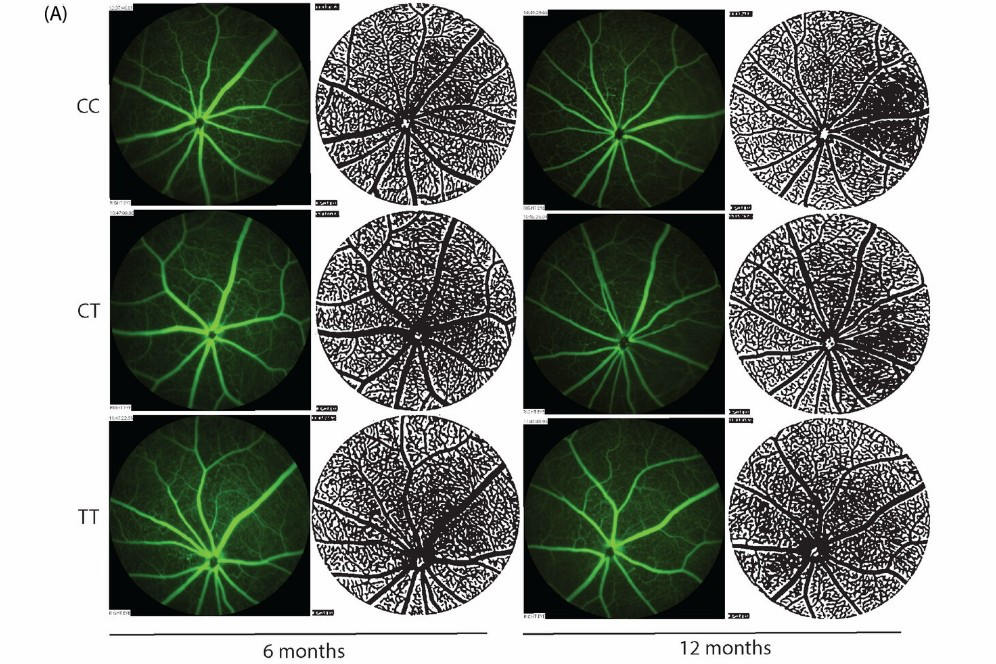
Mice carrying the Mthfr677C>T (CT) mutation, a risk factor for ADRD found in around 40% of humans, exhibit vascular changes in the brain by six months of age, said researchers from The Jackson Laboratory, Bar Harbor, Maine. Unfortunately, decreased retinal vessel density appeared only in 12-month-old mice, challenging retinal vascular density’s usefulness as a biomarker for ADRD, they said. However, instances of retinal arteriovenous crossing and vessel tortuosity were observed in CT mice at six months compared with controls, suggesting these may be useful biomarkers to improve early diagnoses of ADRD, said authors.
Further, the brains and retinas of CT mice were also found to share Alzheimer's disease-relevant differentially expressed proteins, they said.
“These data suggest that assessing age and genetic-driven changes within retinal vasculature represents a minimally invasive method to predict Alzheimer’s disease-related cerebrovascular damage,” they concluded.
The study was published in Alzheimer’s & Dementia.