


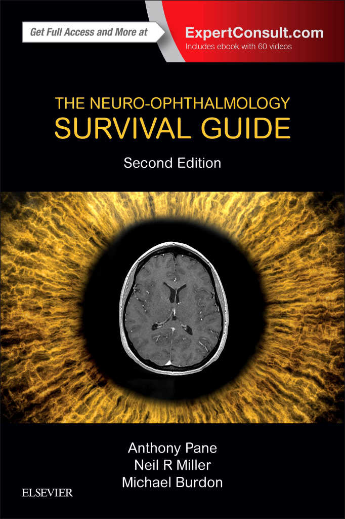
By Anthony Pane, Neil R. Miller and Mike Burdon
Reviewed by Dr Stephen Best*
I was delighted to be invited by the NZ Optics’ editorial team to review this book as I had not taken the opportunity to read the previous edition, although I had heard many references to it from colleagues both here, in New Zealand, and in Australia, where the principle author, Dr Anthony Pane is based.
I have listened to Anthony’s presentations over the years and appreciate his directness about the potential pitfalls (sometimes known as medico-legal watch cases) of neuro-ophthalmic conditions that might have irreversible sight-threatening sequelae or be life-threatening emergencies seen in routine ophthalmic clinics, but potentially under-diagnosed! Additionally, I have spent time with, and greatly respect, Drs Neil Miller and Michael Burdon, so anticipated this small text book would be a good read and live up to expectations!
I was not disappointed; especially after reading the first chapter ‘Staying out of trouble’ which lists 20 neuro “rules” to keep you out of strife. Each rule is illustrated with a case example and cross-referenced to the expanded discussion on that topic in subsequent chapters. This chapter alone should pique interest about common neuro-ophthalmic conditions and, as stated in the introduction, you can’t avoid neuro-ophthalmology - neuro-ophthalmology is special, you want your patients to see well, you want your patients to stay healthy, you want to stay out of trouble, you want to pass your exam (if you still have it ahead of you).
This is a short text book designed to be of everyday practical use for ophthalmologists, trainees, optometrists and neurologists, based on clinical symptoms, examination checklists, management flowcharts and referral guidelines.
There are 12 chapters (340 pages) that cover blurred vision or field loss, swollen disc(s), double vision, unequal pupils and unexplained eye pain, orbital pain or headaches. The final chapter outlines neurophthalmic history and examination, with particular reference to giant cell arteritis and an excellent table 13.1 titled ‘Localizing value of visual field defects’. This last chapter, in my opinion, should be a must read for all ophthalmic trainees not only to reinforce sound clinical practice but also to help with challenging formal examinations.
The chapter on ‘Double vision’ is a wonderful synthesis of an extremely complex topic, but presented in an easily understandable and clinically significant format. While the discussion on cranial mono-neuropathies and localising value flowcharts should guide clinicians to appropriate investigations in particular third nerve disease processes that might have very serious consequences!
One of my favourite chapters, however, dealt with ‘Seeing things’. Patients may see things because of eye, optic nerve or brain disease and unusual visual symptoms need to be explained. Visual illusions and hallucinations are explained with excellent cartoons and flow charts, and Anthony reminds the reader that if the patient presents with visual phenomena that are not consistent with visible intraocular disease processes then referral to a neuro-ophthalmologist or a neurologist is appropriate with neuro-imaging to check for serious brain disease.
This is a great book to have close to your consulting room and, as with many modern texts, when you purchase the second edition it comes with an eBook version which is downloadable to your electronic screen ensuring great portability and a fantastic set of clinical videos.
Dr Stephen Best is a consultant ophthalmologist, with sub-specialities in glaucoma and neuro-ophthalmology, with Auckland Eye and the Greenlane Clinical Centre in New Zealand.