Allergic eye disease: itching to know more?
Allergic conjunctivitis is a common condition affecting approximately 30 to 40% of the population, depending on location.1-3 In the last decade there have been reports of significant increases in all types of allergy, including ocular allergies.1,2 Allergic conjunctivitis can range from mildly irritating to debilitating and potentially vision threatening. It is an overarching label that includes the disease entities of seasonal allergic conjunctivitis (SAC), perennial allergic conjunctivitis (PAC), vernal keratoconjunctivis (VKC), and atopic keratoconjuctivits (AKC).1 Symptoms of allergic eye disease are hyperemia, pruritus (itching), chemosis and lid swelling, with symptoms often closely correlated to the severity of allergy.
Seasonal and perennial allergic conjunctivitis
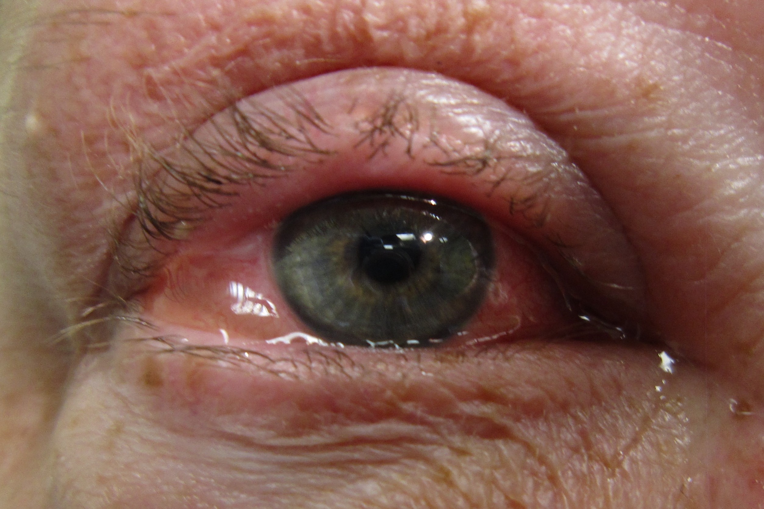
SAC and PAC are the most common presentation of ocular allergies, with estimates of this affecting approximately 15% of the population.1 Affected individuals present with ocular itching, redness, conjunctival papillae and chemosis. The chemosis is often more significant than the ocular hyperaemia, see Fig 1. Papillae can range from small, fine, and velvet-like in appearance (see Fig 2), to larger more defined papillae. Corneal involvement in either SAC or PAC is rare.1
SAC and PAC are both allergen-induced IgE-mediated hypersensitivity reactions. The IgE then binds to, and activates, mast cells inducing the release of histamine, prostaglandins, tryptase, and leukotrienes.4 This results in the early response, which lasts approximately half an hour. This is followed by the late stage reaction, which is due to the mast cell degranulation activating vascular endothelial cells in chemokine and adhesion molecule expressions, resulting in recruitment of inflammatory cells in the conjunctival mucosa and thus, the ongoing reaction when the allergen is no longer present.4 SAC and PAC have the same signs and symptoms due to the underlying similarity of their pathophysiology, however, the allergens causing SAC and PAC vary.1 SAC occurs seasonally and is usually in response to an airborne pollen, with the most common time of occurrence being spring and summer. There are limited reports in winter. While PAC occurs year round to persistently present allergens such as dust mites or animal dander.1
Allergic conjunctivitis is treated in a step-wise approach as documented in Table 1 (below).

Table 1 – Ocular Treatment algorithm for allergic conjunctivitis
Vernal keratoconjunctivitis
VKC is a chronic bilateral allergic inflammation of the ocular surface. It is a non-specific hyperactivity with skin and serum IgE tests to common allergies often being negative. The complex pathogenesis is primarily mediated by Th2-lymphocytes.4 VKC can present in three clinical appearances: limbal, palpebral, and combination. VKC predominantly affects young males and is more common in warm climates during the summer months. Individuals with VKC present with severe ocular itch, photophobia, redness, conjunctival swelling and mucus discharge.1 The characteristic clinical sign is giant papillae on the upper tarsal conjunctiva, sometimes described as ‘cobblestones’, see Fig 3.5

Fig 2. Fine, low grade papillae, with velvet-like appearance

Fig 3. Cobblestone giant papillae
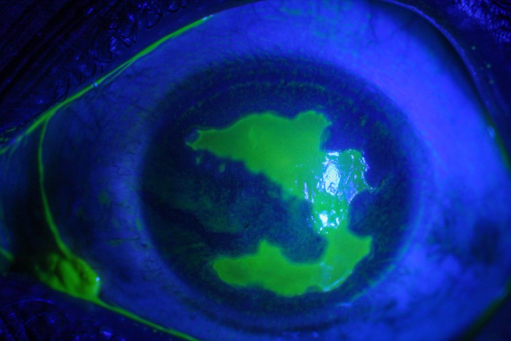
Eosinophils infiltrate and activate in the conjunctiva. They release epitheliotoxic factors that affect the cornea. Initially this presents as punctate keratitis. These can amalgamate, leading to central opaque plaques, and shield ulcers.1 Both are visually threatening and require urgent management. Tantras dots - small distinct opaque clumps with a gelatinous appearance - can be seen on the limbus. Tantras dots tend to be present during active stages of VKC but settle as symptoms subside.1
VKC follows a similar stepwise approach to treatment as SAC and PAC. It can be managed with mast cell stabilisers with, or without, steroids during acute episodes. However, the main challenge is trying to prevent recurrences and the consequences of these, such as shield ulcers and plaques. In cases where mast cell stabilisers and steroids are insufficient options, subtarsal injections of corticosteroids, cyclosporine, tacrolimus or other immunomodulators can be successfully used.5 Surgery can be used to manage giant papillae with shave excision, or to manage shield ulcers with amniotic membrane transplants.5 VKC tends to resolve by adulthood, therefore, appropriate management during childhood and adolescence is essential.
VKC has a large impact on productivity, concentration, and potential for permanent visual loss. Suspected cases of VKC should be referred for ophthalmological management, to prevent any unnecessary visual loss and minimise impact on quality of life.
Atopic keratoconjunctivitis
AKC is considered the ocular form of atopic eczema or dermatitis. It is a bilateral chronic inflammatory disease affecting both the ocular surface and eyelids. It is caused by a chronic degranulation of mast cells and cytokines derived from both Th1- and Th2-lymphocytes.1,4 Skin lesions with an eczematous appearance are found on or around the eyelids. They are often itchy and become worse if scratched. Conjunctival hyperaemia and chemosis can range from severe to mild. Giant papillae and tantras dots may or may not be present.1 AKC is associated with early onset cataracts, regardless of corticosteroid use. AKC and VKC have some overlap, except VKC resolves after the teen years, while AKC can be a lifelong affliction.
Treatment for AKC is dependent on signs and symptoms, with regular monitoring required for cataract development. Again, a stepwise approach should be used to approach treatment for maximum results while minimising risks of side effects.
Allergies in children
Children may potentially have higher rates of ocular allergies than adults.3 Children with atopic co-morbidities including asthma, allergic rhinitis, and atopic dermatitis are most commonly affected.3 Individuals with ocular allergies often report that their eyes are itchy, or they frequently rub their eyes. Often when children are asked, they won’t describe the eye discomfort from allergies as ‘itchy’; they may report blurring, discomfort, or stingy eyes. It is important to ask not only the child about their eye comfort, but also their parents or guardians. Children often eye rub subconsciously, so asking for another’s observations of eye rubbing can give further insight. However, other children choose less conventional ways to cope with the pruritus they are experiencing; this can include eye rolling and excessive blinking. Therefore, a negative response to questions on ocular itch in a child does not preclude ocular allergy.
Interestingly, children with attention deficit hyperactivity disorder (ADHD) have a much higher rate of allergic conjunctivitis than an age-matched control group.3 Allergic conjunctivitis has also been associated with decreased concentration and productivity. Therefore, treatment of allergic eye disease not only relieves symptoms but can bring potential benefits to work, education and focus. For children with attention challenges, such as ADHD, removing other barriers to focus are of great importance.
Clinical tips for allergic conjunctivitis
- Take an accurate history of allergies and family history of atopic disease.
- Ask specific questions about eye rubbing, rolling or abnormal blinking behaviours.
- Undertake a thorough slitlamp examination including:
- Cornea – check for clarity and staining
- Conjunctiva – palpebral and bulbar
- Evert inferior and superior lids to detect papillae
- Document findings with anterior photography if available
- Treat ocular allergy when detected in those who are symptomatic or in younger children when ocular signs are present.
- Follow up patients in six weeks to determine improvement of allergies with treatment.
- In children, ask about focus, improvement at school, and any behavioural changes in eye movements or blinking.
- If AKC or VKC are suspected, refer for further assessment. Co-management can then occur in appropriate cases.
Treating ocular allergies, particularly in children, can result in great improvements in quality of life. This is important for the patient and rewarding for the clinician.
References
- Bielory L, Meltzer EO, Nichols KK, Melton R, Thomas RK, Bartlett JD. An algorithm for the management of allergic conjunctivitis. InAllergy and asthma proceedings 2013 Sep 1 (Vol. 34, No. 5, pp. 408-420). OceanSide Publications, Inc.
- La Rosa M, Lionetti E, Reibaldi M, Russo A, Longo A, Leonardi S, Tomarchio S, Avitabile T, Reibaldi A. Allergic conjunctivitis: a comprehensive review of the literature. Italian journal of pediatrics. 2013 Dec 1;39(1):18.
- Dupuis P, Prokopich CL, Hynes A, Kim H. A contemporary look at allergic conjunctivitis. Allergy, Asthma & Clinical Immunology. 2020 Dec 1;16(1):5.
- Sánchez-Hernández MC, Montero J, Rondon C, del Castillo Benitez JM, Velázquez E, Herreras JM, Fernández-Parra B, Merayo-Lloves J, Del AC, Vega F, Valero A. Consensus document on allergic conjunctivitis (DECA). Journal of investigational allergology & clinical immunology. 2015;25(2):94-106.
- Singhal D, Sahay P, Maharana PK, Raj N, Sharma N, Titiyal JS. Vernal keratoconjunctivitis. Survey of ophthalmology. 2019 May 1;64(3):289-311.

Dr Samantha Simkin is a therapeutically qualified optometrist working at Mortimer Hirst Optometrists, a member of the BLENNZ National Assessment Team and a Research Fellow in the Department of Ophthalmology at the University of Auckland.


