Iris melanoma: a review
Iris naevi are common, making up to 63% of melanocytic iris tumours found in adults1. In contrast, iris melanomas are rare, making up only 2-8% of all uveal melanomas2. Most are believed to arise from iris naevi, with an estimated transformation rate of 4% over 10 years, and 11% over 20 years3. Iris melanoma typically display two patterns of growth, circumscribed or diffuse. Progress tends to be slow - either locally along the surface of the iris, locally into the anterior chamber or invading into the ciliary body4. Metastases of iris melanoma occurs in up to 10% of cases over 20 years and are influenced by factors such as the cytology of the tumour, increased age of the patient and angle involvement. This renders the distinction between naevus and melanoma, and then the different histological subtypes, clinically important.
Iris naevi- ‘The ABCDEF Guide’3
Certain clinical features of iris naevi have been identified as indicators of increased risk for transformation into iris melanoma. A study done by Shields et al, which analysed 1611 consecutive patients with an initial diagnosis of iris naevus, developed ‘The ABCDEF Guide’ - a mnemonic for remembering factors predictive of transformation: A - age < 40 years, B - blood (hyphaema), C - clock hour inferior, D - diffuse, E - ectropion uveae, F - feathery margin3. While the mnemonic is helpful, important features are missing such as large size, nodule formation, elevated intra-ocular pressure and prominent intralesional vasculature5.
The use of sequential photographs via slit lamp photography is extremely helpful for monitoring the size and growth of iris lesions. The frequency at which these photographs should occur depends on if there are features present which make the naevi higher risk and whether there is a history of suspected recent change. Should any changes in naevi be noticed, referral for ophthalmic review is advised. Just as patients are educated to monitor their cutaneous naevi, they should also be encouraged to monitor their iris naevi for any obvious changes if possible.
Epidemiology of iris melanoma
The prevalence of iris melanoma is increasing6. The estimated incidence of iris melanoma in New Zealand is 0.09/100,000 per year6, which is slightly higher than the estimated European incidence (0.02-0.08/100,000 per year)7. This may be attributed to the higher amount of solar radiation experienced in New Zealand. The superior part of the iris, which is better protected from UV radiation by the upper eyelid, rarely experiences iris melanoma6.
The average age of patients with iris melanoma is 64 years of age8, with an equal distribution between male and females9. Risk factors include pre-existing iris naevi, fair skin with cutaneous naevi and non-brown coloured irises8. One study found 4% of patients diagnosed with iris melanoma had a history of skin melanoma10.
Presentation
The most common presentation of iris melanoma is the sudden rapid growth of a previously existing pigmented iris lesion. Most of these tend to be brown in colour (65.6%), but also exist as amelanotic (9.9%) or multi-coloured (9,9%)8. The shape is most commonly nodular (78.4%) as opposed to flat (21.6%) and existing in the inferior clock face (78.4%)9. Common associated features include a corectopia (the drawing of the pupil from its usual central position, 62%), ectropion uveae (44%), intrinsic tumour vessels (43%), hyphaema (9%) secondary glaucoma and tumour induced cataract (14%)5.
Histology
Iris melanomas can be divided into three broad histological categories: spindle- cell, epithelioid-cell and mixed – with spindle-cell tumours being the most common and carrying the best prognosis9. Mixed or epithelioid tumours have been found to have an estimated eight times higher risk of mortality8.
Management of iris melanoma
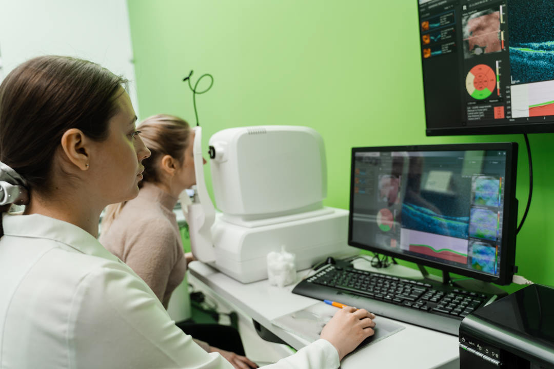
The course of management for iris melanoma depends on many factors, including the size of the tumour, invasion into the anterior chamber angle and the presence of melanoma-related complications such as raised intraocular pressure. An initial thorough inspection of the lesion is recommended, followed by an investigation for involvement of surrounding structures, including the anterior chamber, angle, lens and cornea5. Diagnostic procedures such as ultrasonographic biomicroscopy and anterior segment optical coherence tomography (OCT) are useful for differentiating between cystic and solid lesions10, as well as assessing the ciliary body for involvement. Ultrasound-guided fine-needle aspiration biopsy to confirm cytology can also play a role in guiding the course of management.
Watchful waiting
Watchful waiting is an option for lesions that are slow growing or have been histologically identified as composed of spindle type-A cells. These melanomas have a very low risk of local invasion and metastasis11.
Surgical Excision
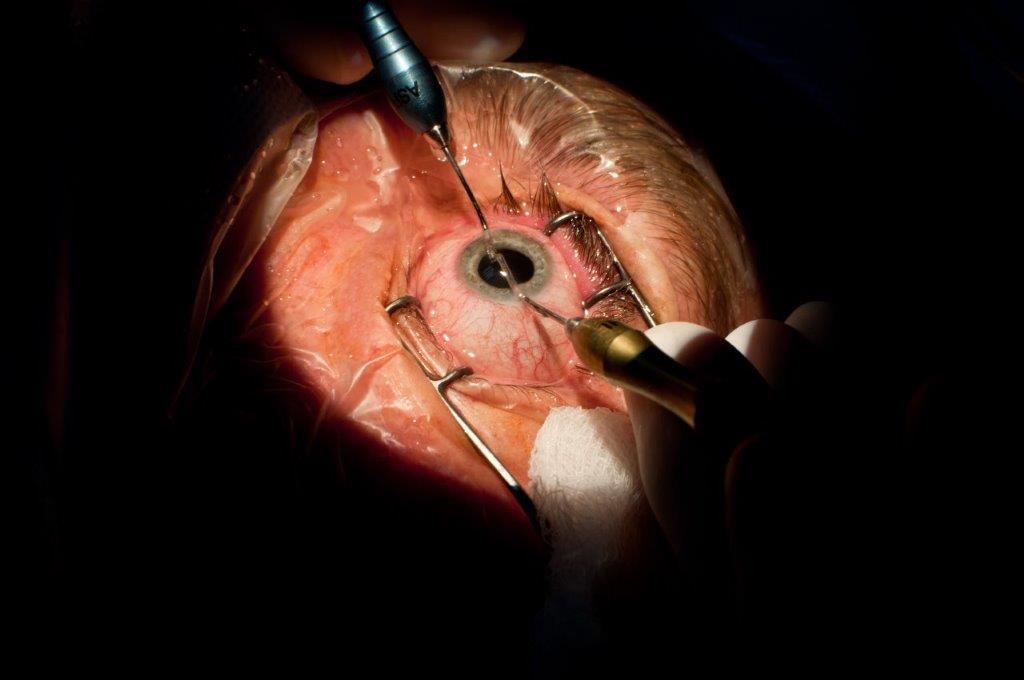
For lesions identified as being localised and without evidence of tumour seeding, local excision via iridectomy or iridocyclectomy is an appropriate choice of management. For these patients, the most common post-operative complaint is glare – experienced in up to 25% of cases12. This can be managed with either a pupil reconstruction or tinted glasses. Other complications include incomplete excision of the lesion, a dislocated lens, cataract progression and post-operative glaucoma4.
Radiotherapy
Radiotherapeutic approaches to iris melanoma include proton beam therapy and plaque radiotherapy. Proton beam therapy involves a highly collimated external beam that uses protons instead of x-rays13. Plaque radiotherapy is a disc of radioactive material (commonly Iodine 125) delivering localised trans-corneal radiotherapy to an iris lesion14. Indications for radiotherapy include residual tumour following surgical excision, tumour recurrence and diffuse or multifocal involvement. Radiation-induced side effects are common. Shields et al reported at five years following treatment, patients developed cataracts (70%), corneal conditions (9%) and neovascular glaucoma (8%)14. An additional complication specific to proton beam therapy is limbal stem cell deficiency12.
A review done by Popovic et al12 comparing the radiotherapeutic and surgical management of iris melanoma found that radiotherapeutic approaches are now being used more frequently (proton beam 49.4%, plaque 31.4%) than surgical resection (19.2%). Other findings included lower rates of local recurrence and metastatic development with radiotherapy (0-5%) than surgery (0-14%, including enucleation). However, surgical management tended to show lower rates of cataract and glaucoma development following treatment and avoids the radiotherapeutic specific complications such as limbal stem cell deficiency12.
Enucleation
Enucleation is reserved as a last resort treatment for tumours that show aggressive recurrence, extensive growth and invasion, poor vision potential and uncontrollably high intraocular pressure with pain12,14.
Summary
There is considerable overlap between the features of iris naevus and melanoma, warranting monitoring of naevi by patients and their families, optometrists and ophthalmologists. The use of radiotherapeutic approaches are becoming more popular, with studies showing lower rates of recurrence and metastases compared to local surgical excision. However, this must be balanced against the higher rates of common complications such as cataract, glaucoma and limbal stem cell deficiency. Given the rarity and heterogeneity of iris melanoma, there remains a need for more research regarding its monitoring and optimum timing for intervention.
Images courtesy of Professor Charles McGhee.

Fig 3. (a) Iris melanoma with visible incisional biopsy site; (b) following excisional biopsy; (c) following phacoemulsification and insertion of an intraocular lens and artificial iris
References:
Shields, Carol L., et al. "Clinical survey of 3680 iris tumors based on patient age at presentation." Ophthalmology2 (2012): 407-414.
- Shields, Carol L., et al. "Clinical spectrum and prognosis of uveal melanoma based on age at presentation in 8,033 cases." Retina7 (2012): 1363-1372.
- Shields, Carol L., et al. "Iris nevus growth into melanoma: analysis of 1611 consecutive eyes: the ABCDEF guide." Ophthalmology4 (2013): 766-772.
- Conway, R. M., et al. "Primary iris melanoma: diagnostic features and outcome of conservative surgical treatment." British journal of ophthalmology7 (2001): 848-854.
- Shields, Carol L., et al. "Iris melanoma: risk factors for metastasis in 169 consecutive patients." Ophthalmology1 (2001): 172-178.
- Michalova, Kira, et al. "Iris melanomas: are they more frequent in New Zealand?." British journal of ophthalmology1 (2001): 4-5.
- McGalliard, J. N., and P. B. Johnston. "A study of iris melanoma in Northern Ireland." British journal of ophthalmology8 (1989): 591-595.
- Khan, Samira, et al. "Clinical and pathologic characteristics of biopsy-proven iris melanoma: a multicenter international study." Archives of Ophthalmology1 (2012): 57-64.
- Jakobiec, Frederick A., and Glenn Silbert. "Are most iris' melanomas' really nevi?: A clinicopathologic study of 189 lesions." Archives of Ophthalmology12 (1981): 2117-2132.
- Pavlin, Charles J., et al. "Ultrasound biomicroscopy of anterior segment tumors." Ophthalmology8 (1992): 1220-1zffwtki.
- Geisse, Lawrence J., and Dennis M. Robertson. "Iris melanomas." American journal of ophthalmology6 (1985): 638-648.
- Popovic, Marko, et al. "Radiotherapeutic and surgical management of iris melanoma: A review." Survey of ophthalmology(2017).
- Rahmi, Ahmed, et al. "Proton beam therapy for presumed and confirmed iris melanomas: a review of 36 cases." Graefe's Archive for Clinical and Experimental Ophthalmology9 (2014): 1515-1521.
- Shields, Carol L., et al. "Iris melanoma management with iodine-125 plaque radiotherapy in 144 patients: impact of melanoma-related glaucoma on outcomes." Ophthalmology1 (2013): 55-61.
- Shields, Carol L., et al. "Custom-designed plaque radiotherapy for nonresectable iris melanoma in 38 patients: tumor control and ocular complications." American journal of ophthalmology5 (2003): 648-656.
About the authors

Amy Clucas

Dr Jennifer Court
*Amy Clucas is a medical student at the University of Auckland who developed an interest in ophthalmology on her two-week attachment last year. She completed a selective in ophthalmology with the Greenlane Clinical Centre’s eye department and says she’s looking forward to future experiences in the field.
Dr Jennifer Court trained in ophthalmology in the UK. She is a cornea and anterior segment fellow at Greenlane Clinical Centre, Auckland.


