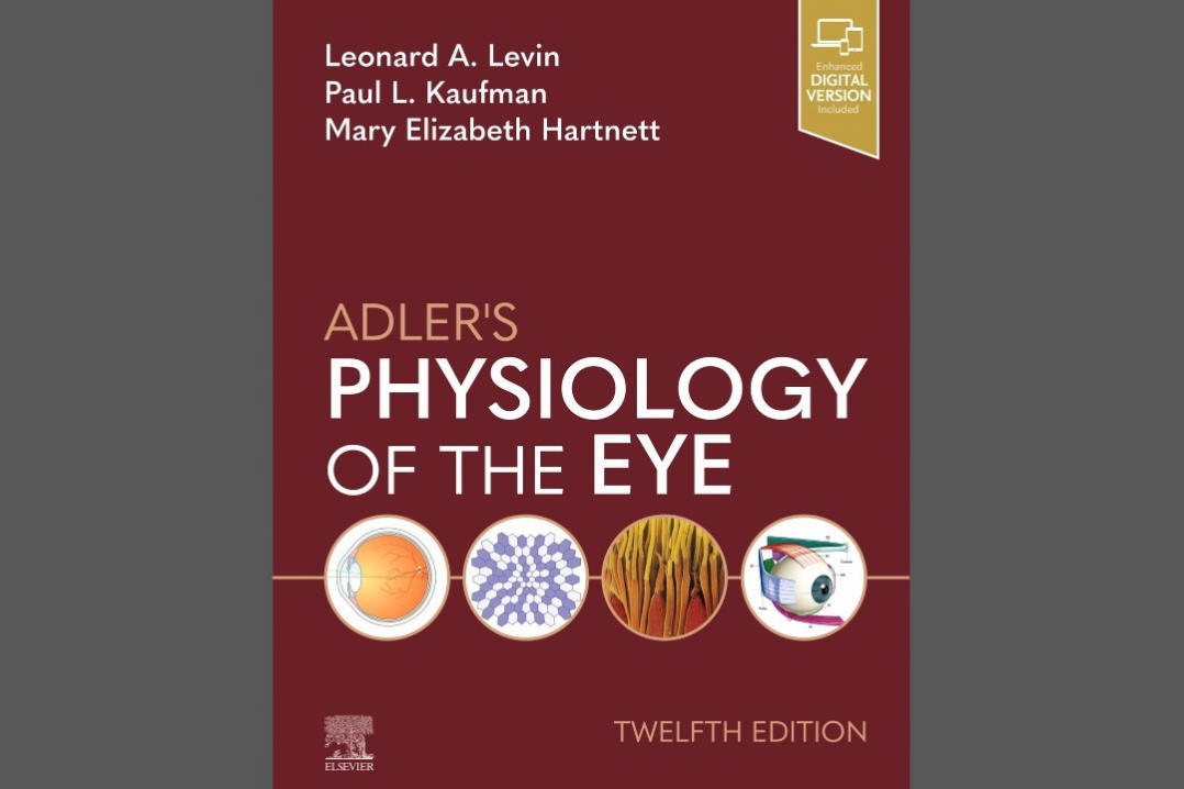


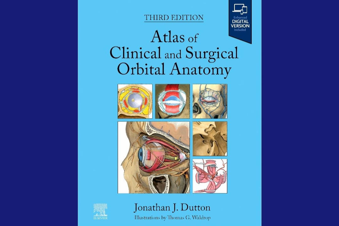
The Atlas of Clinical and Surgical Orbital Anatomy (3rd edition) is an invaluable guide for orbital surgeons and those who work closely with the orbital region, such as ENT and maxillofacial surgeons.
In this third edition, Professor Emeritus Jonathan Dutton, from the University of North Carolina, US, provides more detailed information on the levator and Müller muscles of the upper lids, as well as the extraocular pulley systems. These insights have revolutionised our understanding of their functionality and continue to challenge and refine our surgical approaches and techniques. He has also added a new chapter on nasal cavity and paranasal sinuses.
Each chapter covers a different system of the eye: Cavernous sinus, Osteology of the orbit, Extraocular muscles, Nerves, Arterial supply, Venous and lymphatic systems, Orbital fat and connective tissue, Eyelids, Lacrimal system, and Nasal cavity and sinuses. This makes reading and accessing information straightforward. Every chapter also includes extensive embryological studies, followed by detailed descriptions of individual structures and their clinical correlations.
The book contains a large collection of medical illustrations. These are simple and concise drawings that effectively help readers conceptualise the anatomy and apply it to real-life scenarios. The illustrations are presented from various angles to help build a three-dimensional image in the reader's mind. The availability of an eBook version also facilitates easier access to these detailed and informative illustrations on mobile devices.
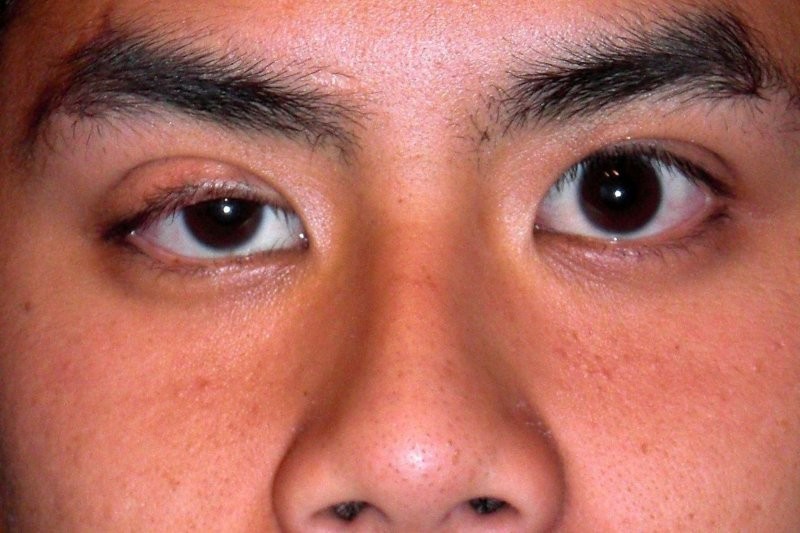
One of the most valuable aspects of this atlas are the extensive measurements drawn from the literature, complete with references. These measurements include: the insertion, position and length of extraocular muscle; various muscles and ligaments of the lids (palpebral levator, Muller's and Whitnall's ligaments); the nasolacrimal duct system, etc. As an oculoplastic surgeon, I found this knowledge particularly helpful for navigating and identifying anatomical structures and their relative positions to adjacent tissues when the surgical view is obscured by blood.
Another significant feature of the atlas are the clinical correlations provided at the end of each chapter. These sections briefly discuss conditions related to the anatomical structure or system, helping readers apply their anatomical knowledge in clinical practice without delving too deeply into pathophysiology and management.
The last two chapters, which cover histology and radiological correlations, are also valuable additions to the atlas. The CT images are well-presented with side-to-side medical illustrations for comparison.
If you are interested in orbital anatomy, this atlas is an excellent resource.

Dr Dickson Wong is a consultant ophthalmologist at Christchurch Hospital and Southern Eye Specialists. Dual trained in glaucoma and oculoplastics, he is the ophthalmology trainee supervisor in Christchurch and a member of the RANZCO selection committee for the New Zealand training network.