US researchers have developed a new protocol to triage potential eye-stroke patients, saying it will flag those at risk of brain stroke and help save vision.
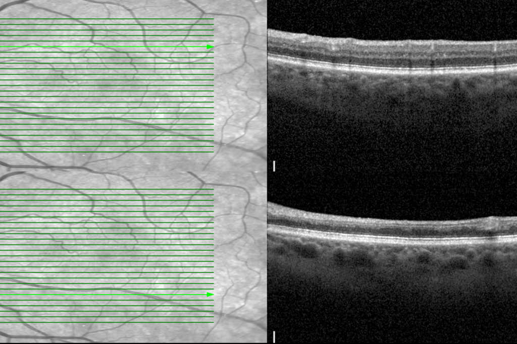
The protocol, developed by researchers at the New York Eye and Ear Infirmary of Mount Sinai (NYEE), uses optical coherence tomography (OCT) when a patient with suspected central retinal artery occlusion enters the medical system. “This reduces the need for onsite ophthalmology consults, which are often not immediately available,” explained Dr Richard Rosen, head of the Retina Service at Mount Sinai. OCT images are then sent to remote, on-call retinal specialists who can make an instant diagnosis. If they confirm an eye stroke diagnosis, the vascular interventional neuroradiologists can then deliver an infusion of tissue plasminogen activator (tPA) into the blocked ophthalmic artery, said the research team.
During the protocol’s first 18 months of use, 42% of 59 patients had a confirmed eye stroke based on OCT imaging and follow-up examination, after receiving treatment around 2.5 hours after arrival. Researchers recorded a statistically significant improvement in these patients’ visual acuity four weeks after tPA treatment, with a “dramatic improvement” in 66% of patients within 24 hours of treatment. Of these, 56% had maintained improved vision one month later. On average, the patients’ vision improved from counting fingers to 20/100, with 44% of those treated with tPA improving to 20/40 or better, which constitutes unrestricted driving vision in New York State, noted authors.