


Researchers in Australia found high-resolution microperimetry performed better than standard automated perimetry (SAP) in detecting glaucomatous damage.
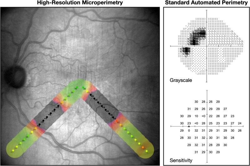
Fundus-tracked perimetry, commonly described as microperimetry, allows real-time compensation for fixation drifts so stimuli can be consistently presented at the same retinal locations, said researchers from the Centre for Eye Research Australia, writing in Clinical & Experimental Ophthalmology. They assessed 250 eyes from 200 individuals using high-resolution microperimetry testing of a hemifield using a stimulus pattern optimised to sample typical arcuate patterns of visual field (VF) loss in glaucoma. SAP was performed using a 24–2 stimulus pattern. The presence of glaucomatous damage in each hemifield was then assessed by two graders. OCT imaging was also performed to obtain three imaging outputs for evaluation of the presence of glaucomatous damage.
High-resolution microperimetry performed significantly better than 24–2 SAP at predicting the presence of OCT-defined glaucomatous damage, said researchers.
These findings suggest high-resolution microperimetry could provide a more effective means for the characterisation of glaucomatous visual field loss, they said. “While this approach may achieve higher performance than a standard 24–2 VF test for the specific task of detection, one of its main disadvantages is its limited ability to directly spatially characterise the entire visual field, which is important for understanding the current and future risk of functional disability,” researchers said.