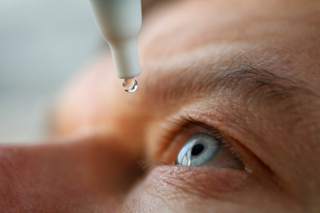


The impact of disease on the ocular surface is often assessed clinically by checking for fluorescein staining of the corneal epithelium, to highlight areas of damage secondary to trauma, infection or desiccation. In dry eye disease, corneal staining is typically superficial, punctate and located inferiorly, as the interpalpebral zone is the area least protected by the eyelids and thus has the most prolonged exposure to the external environment.
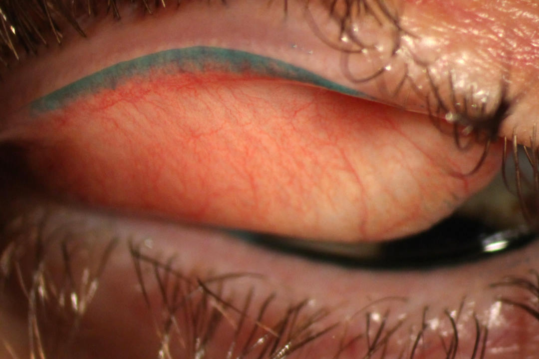
Previous research from the Ocular Surface Laboratory (OSL) has shown that corneal staining tends to occur late in the natural history of dry eye development, often not until patients are in their 50s¹. This late-stage finding is thus often observable only once dry eye has been present for many decades and has become more severe in nature. In contrast, lid-wiper epitheliopathy (LWE) is a marker of epithelial damage at the eyelid margin’s ‘lid-wiper zone’ – the area in constant contact with the ocular surface during blinking – and this has been shown to be visible decades earlier in the disease process¹, potentially making it a superior marker of dry eye, especially in more mild disease².
Most recently, the OSL team has published an article that explores the diagnostic performance of different types of ocular surface staining in detecting dry eye, according to the global consensus TFOS DEWS II diagnostic criteria, to help clinicians choose the most appropriate clinical diagnostic tests to apply³. These findings are based on data from 2066 individuals, with or without dry eye, who agreed to undergo a full dry eye workup, involving both symptoms and signs assessment, and contribute their data to our local research registry.
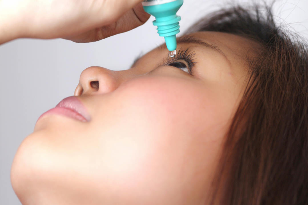
Masked grading of corneal, conjunctival and lid margin fluorescein and lissamine green staining was undertaken by an independent grader, against two established scoring schemes: the Sjögren's International Collaborative Clinical Alliance (SICCA) corneal and conjunctival staining scoring and Korb lid wiper epitheliopathy (LWE) grading.
Overall, 39% of the participants met the criteria for dry eye disease diagnosis according to TFOS DEWS II, with 9% classified as having moderate-to-severe disease. It was observed that the discriminative ability of LWE was significantly better than that of either corneal or conjunctival staining. The optimal thresholds for the SICCA corneal and conjunctival staining scores to detect dry eye were each ≥1 (at least one dot of corneal fluorescein staining or at least 10 dots of conjunctival lissamine green staining), and for the Korb LWE grading was ≥1 (lissamine green staining of at least 2mm in length and 25% in width). LWE was observed more commonly than either corneal or conjunctival staining across both disease severity groups and correlated more consistently with the other measured ocular surface parameters.
This study confirms that LWE is more useful diagnostically than corneal and conjunctival staining in detecting dry eye and offers scientific evidence supporting the routine inclusion of LWE assessment as part of a dry eye disease diagnostic workup.
References
1. Wang MTM, Muntz A, Lim, J, Kim JS, Lacerda L, Arora A, Craig JP. Ageing and the natural history of dry eye disease: a prospective registry-based cross-sectional study Ocul Surf. 2020 Aug 3;18(4): 736-741.
2. Wang MTM, Dean SJ, Xue AL, Craig JP. Comparative performance of lid wiper epitheliopathy and corneal staining in detecting dry eye disease. Clin Exp Ophthalmol. 2019;47(4):546-548.
3. Wang MTM, Power B, Xue AL, Craig JP. Discriminative performance of ocular surface staining and lid wiper epitheliopathy in dry eye disease: an investigator-masked, prospective registry-based, diagnostic accuracy study. Ocul Surf. 2024; 34:165-172

Professor Jennifer Craig heads the Ocular Surface Laboratory within the Department of Ophthalmology at the University of Auckland.