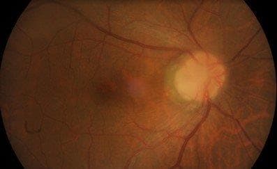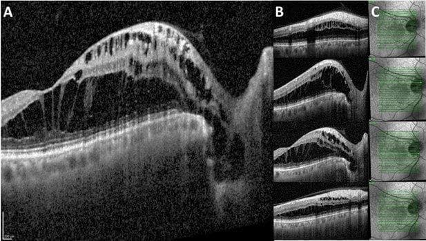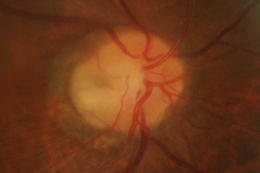
A 58-year-old male with no underlying medical history was referred for retinal oedema adjacent to the optic nerve head with cystic changes and a shiny appearance on the overlying retina for the right eye. Unaided visual acuity was 6/9 and 6/6. He was asymptomatic and was incidentally noted to have macula oedema during a routine examination. The anterior segment examination was normal, but the right eye optic disc assessment showed a greyish-white depressed area in the temporal quadrant (Figs 1 and 2) along with cystic changes at the macula. The left eye examination was normal. OCT examination of the right eye showed schisis-like separation of retinal layers, most marked in the outer retinal layers, extending up to the optic disc temporal margins (Fig 3). There appeared to be a clear communication between the optic disc and the schisis-like separation of outer retinal layers.