Quick scan detects early retinal damage
The developers of flicker optoretinography (f-ORG), which allows visualisation of molecular-level elongations in photoreceptors, said the technique can be used to assess vision loss and neurodegeneration and evaluate potential retinal therapies by detecting pathological changes much earlier than traditional methods.
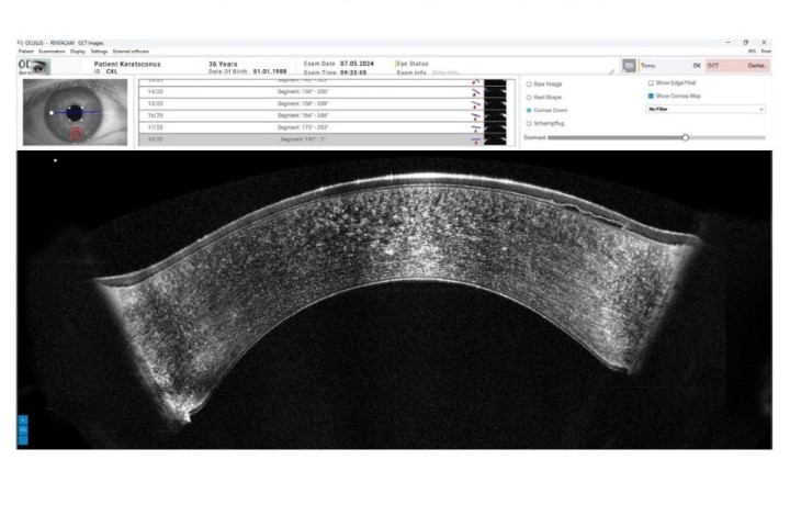
Researchers from Poland’s International Centre for Eye Research described f-ORG as a non-invasive, contact-free, fast and quantitative technology. The technique uses spatial-temporal optical tomography OCT to perform about 200 three-dimensional scans per second, which allows real-time observation of lengthening or shortening of the cone outer segment (COS) in response to light. Changes in COS length reflect the functional activity and health of the retina and result from conformational changes in phosphodiesterase 6 (PDE6), a key enzyme in phototransduction, said researchers. To identify the processes responsible for changes in photoreceptor length, they used sildenafil (Viagra), a known inhibitor of PDE6, which reduced the elongation of rods in a live mouse model.
“Our method enables tracking of molecular mechanisms of phototransduction without the need for prolonged exposure to darkness and without contact with the surface of the eye. This is a significant step forward in the diagnosis of retinal diseases,” said study co-author Professor Maciej Wojtkowski.
The study is published in Proceedings of the National Academy of Sciences.


