Cells of the retinal pigment epithelium (RPE) form unique patterns that can be used to track changes in the back of the eye, researchers at the US National Eye Institute (NEI) have found.
Using a combination of adaptive optics imaging and a fluorescent dye, the researchers used the RPE patterns to track individual cells in healthy volunteers and people with retinal disease. The new finding could provide a way to study the progression and treatment of blinding diseases that affect the RPE.
“Studying cells of the retinal pigment epithelium in the clinic is like looking into a black box. RPE cells are difficult to see, and by the time signs of disease are detectable with conventional techniques, a lot of damage has often already occurred,” said lead author Dr Johnny Tam. “This study is proof-of-concept that we can use a fluorescent dye to reveal this unique fingerprint of the RPE, and to monitor the tissue over time.”
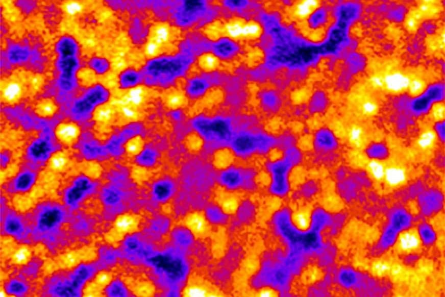
The RPE is a cell layer that lies next to and maintains the health of the retina’s light-sensing photoreceptors. Because the cells contain pigment, and thus absorb incoming light, the thin layer of RPE tissue is difficult to image. Tam used an FDA-approved fluorescent dye called indocyanine green (ICG). While it fades from the blood vessels within about thirty minutes, the dye persists in the RPE for several hours, revealing a fluorescent mosaic pattern, with some cells appearing brighter and others dimmer.
“Initially, we didn’t know how the dye was going to look,” said Tam. “We put the dye in and we got this pattern that at first looked kind of random. It was a big surprise that we could come back after a year, re-inject the dye, and see the same pattern.”