


Hamilton-based vitreoretinal specialist Dr Stephen Guest explains how Optos’ ultra-widefield imaging technology is making a difference to his practice and his patients. By Lesley Springall for Optos.

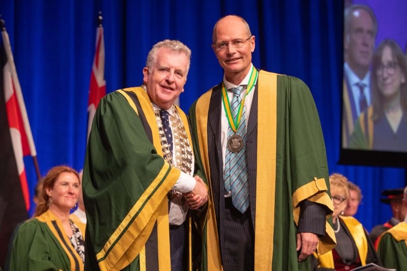
Dr Stephen Guest
Why did you decide to introduce Optomap ultra-widefield retinal imaging into both your private and public practice?
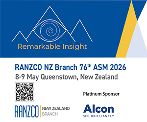
Photo-documenting peripheral fundal lesions, both in colour and with angiography, was often challenging prior to having the Optos. Now it's easy to perform and doesn't require a lot of training.
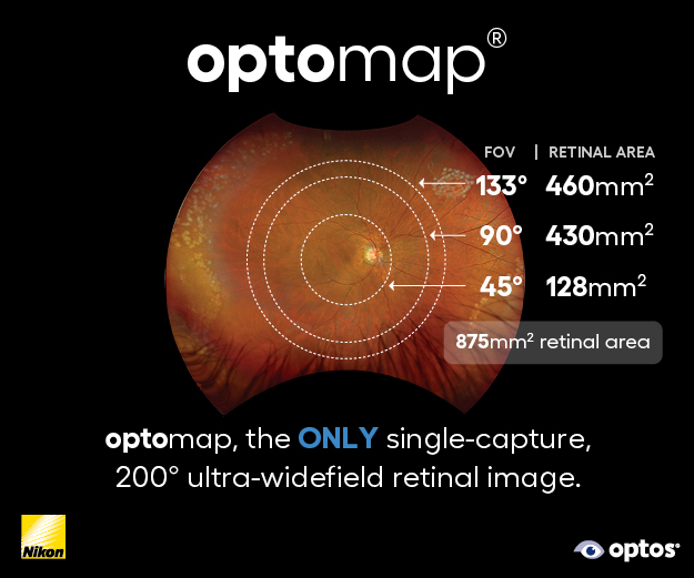
The Optos captures the horizontal meridian better than any other widely available technologies. The ability to perform widefield (and particularly temporal) fluorescein/ICG angiography is very useful for characterising some of the more unusual conditions, such as retinal vasculitis, FEVR (familial exudative vitreoretinopathy), PEHCR (peripheral exudative haemorrhagic chorioretinopathy) and Coats disease, where you need to know what's happening in that far periphery.
The Silverstone Optos OCT facility is particularly useful for diabetic screening. The colour photos are sensitive at picking up proliferative disease and you often see more of the fundus with the Optos than you do with a normal exam. Taking a macular OCT in patients with diabetes at the same time as a widefield colour photo, gives you almost all the information you need regarding the diabetic retinopathy status of that patient.
With choroidal naevi, the Optos peripheral OCT capability gives information on the height of the lesion and whether the naevus has subretinal fluid or not, which can be a sign of potential malignant transformation. Differentiating a retinoschisis from a chronic atrophic peripheral retinal detachment is another useful OCT application, guiding your decision as to whether observation or active management is required.
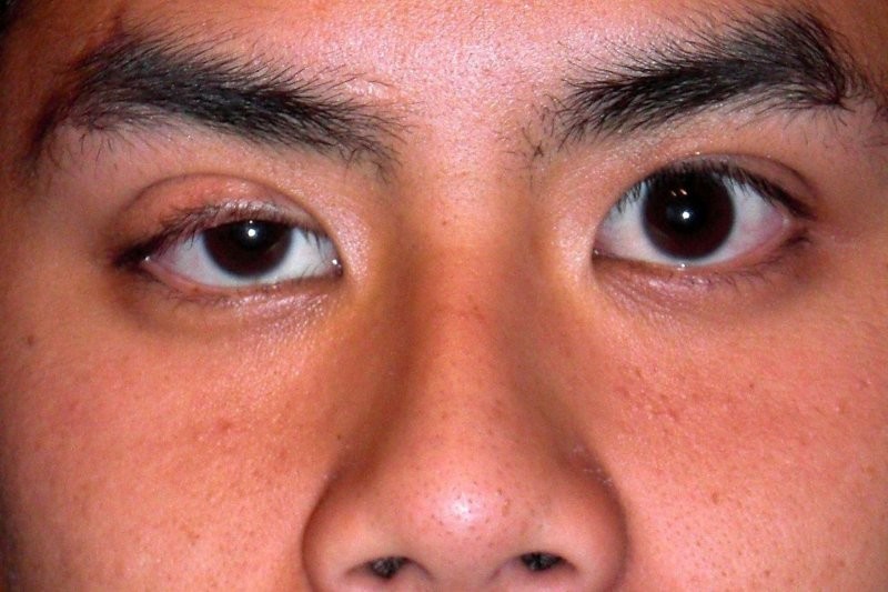
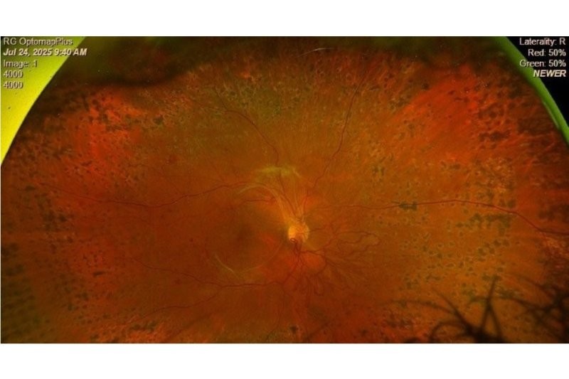
A peripheral exudative haemorrhagic chorioretinopathy lesion in an 80-year-old. Optos Silverstone’s image “helps confirm the lesion is not a choroidal mass but instead a subretinal/subRPE haemorrhage,” says Dr Stephen Guest
What impact do you think the Optos Silverstone has had on reducing the need for multiple imaging devices or repeat appointments?
In the public health service, the Silverstone has made a big difference. We have a three-tier system whereby in the first instance, all diabetics have regular 1-2 yearly non-mydriatic photoscreening in the community. If there are concerns regarding possible diabetic macular oedema or worrisome retinopathy, they are then referred to a Silverstone Optos, nurse-led ‘virtual’ clinic. Here they undergo widefield Optos colour photography and have a macular OCT all on the same machine. Generally the patients then stay in the virtual clinic until they require treatment of some sort, when they will be referred to a doctor-led diabetic eye clinic. Although the virtual clinic images need assessment, this second-tier clinic reduces the need for diabetic patients to have face-to-face meeting with the ophthalmologist – a limited resource in our current environment.
What else would you say to other eyecare professionals who may be considering investing in Optomap?
Just that they’ll find far more uses than they anticipate with this technology… because of that amazing field of view.
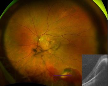
Silverstone RGB, Optos’ pathology reference guide
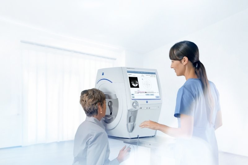
Combining 200° single shot true-to-life colour imaging with advanced swept-source OCT, the Silverstone RGB delivers nine imaging modalities in a single device, enabling clinicians to capture, visualise and analyse pathology across every layer of the retina. The nine modalities include optomap colour RGB for true-to-life retinal colour imaging; optomap colour RG as the proven clinical standard, supported by more than 3,000 peer-reviewed studies; optomap fluorescein angiography for vascular evaluation and disease detection; optomap indocyanine green angiography for enhanced choroidal and vascular imaging; optomap sensory red-free for highlighting nerve fibre and vascular structures; optomap choroidal imaging for deep tissue visualisation; optomap green autofluorescence for identifying RPE changes and metabolic activity; optomap blue autofluorescence for visualising subtle retinal pathology; and swept-source OCT for high-resolution, navigable imaging anywhere in the retina.
Optomap Recognising Pathology is a searchable reference resource to support clinical decision-making and to assist healthcare professionals identify pathology in optomap retinal images and OCT scans. All case images are from the latest ultra-widefield optomap device technology with results available by pathology and/or optomap image modality. For more visit: https://www.optos.com/recognizing-pathology/.