Hamilton-based vitreoretinal specialist Dr Stephen Guest explains how Optos’ ultra-widefield imaging technology is making a difference to his practice and his patients. By Lesley Springall for Optos.

Dr Stephen Guest
Why did you decide to introduce Optomap ultra-widefield retinal imaging into both your private and public practice?
Photo-documenting peripheral fundal lesions, both in colour and with angiography, was often challenging prior to having the Optos. Now it's easy to perform and doesn't require a lot of training.
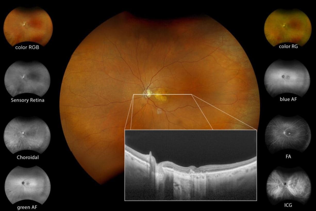
The Optos captures the horizontal meridian better than any other widely available technologies. The ability to perform widefield (and particularly temporal) fluorescein/ICG angiography is very useful for characterising some of the more unusual conditions, such as retinal vasculitis, FEVR (familial exudative vitreoretinopathy), PEHCR (peripheral exudative haemorrhagic chorioretinopathy) and Coats disease, where you need to know what's happening in that far periphery.
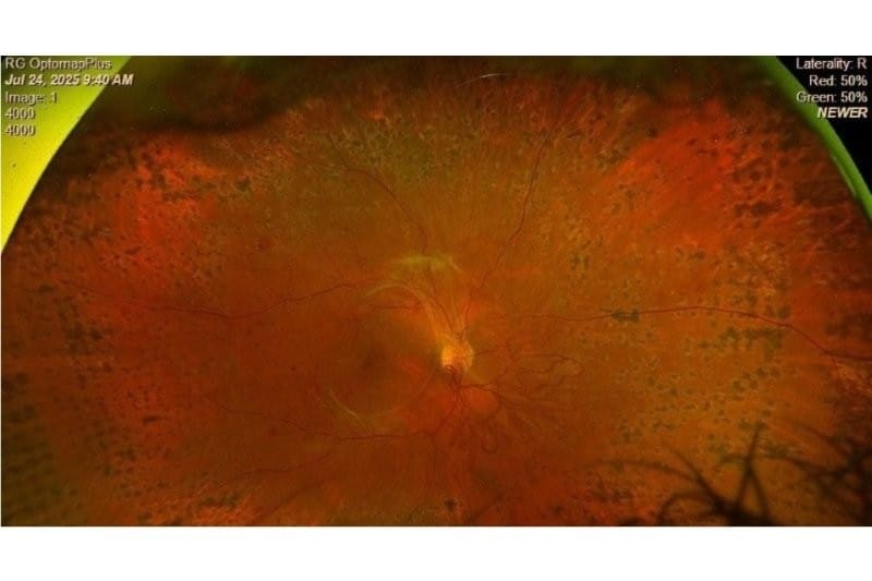
The Silverstone Optos OCT facility is particularly useful for diabetic screening. The colour photos are sensitive at picking up proliferative disease and you often see more of the fundus with the Optos than you do with a normal exam. Taking a macular OCT in patients with diabetes at the same time as a widefield colour photo, gives you almost all the information you need regarding the diabetic retinopathy status of that patient.
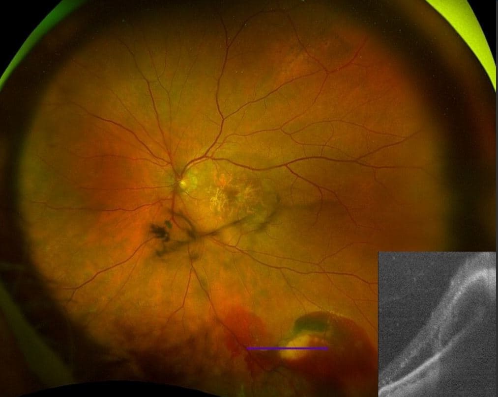
With choroidal naevi, the Optos peripheral OCT capability gives information on the height of the lesion and whether the naevus has subretinal fluid or not, which can be a sign of potential malignant transformation. Differentiating a retinoschisis from a chronic atrophic peripheral retinal detachment is another useful OCT application, guiding your decision as to whether observation or active management is required.
What impact do you think the Optos Silverstone has had on reducing the need for multiple imaging devices or repeat appointments?